Copyright
©The Author(s) 2021.
World J Clin Cases. Mar 16, 2021; 9(8): 2015-2021
Published online Mar 16, 2021. doi: 10.12998/wjcc.v9.i8.2015
Published online Mar 16, 2021. doi: 10.12998/wjcc.v9.i8.2015
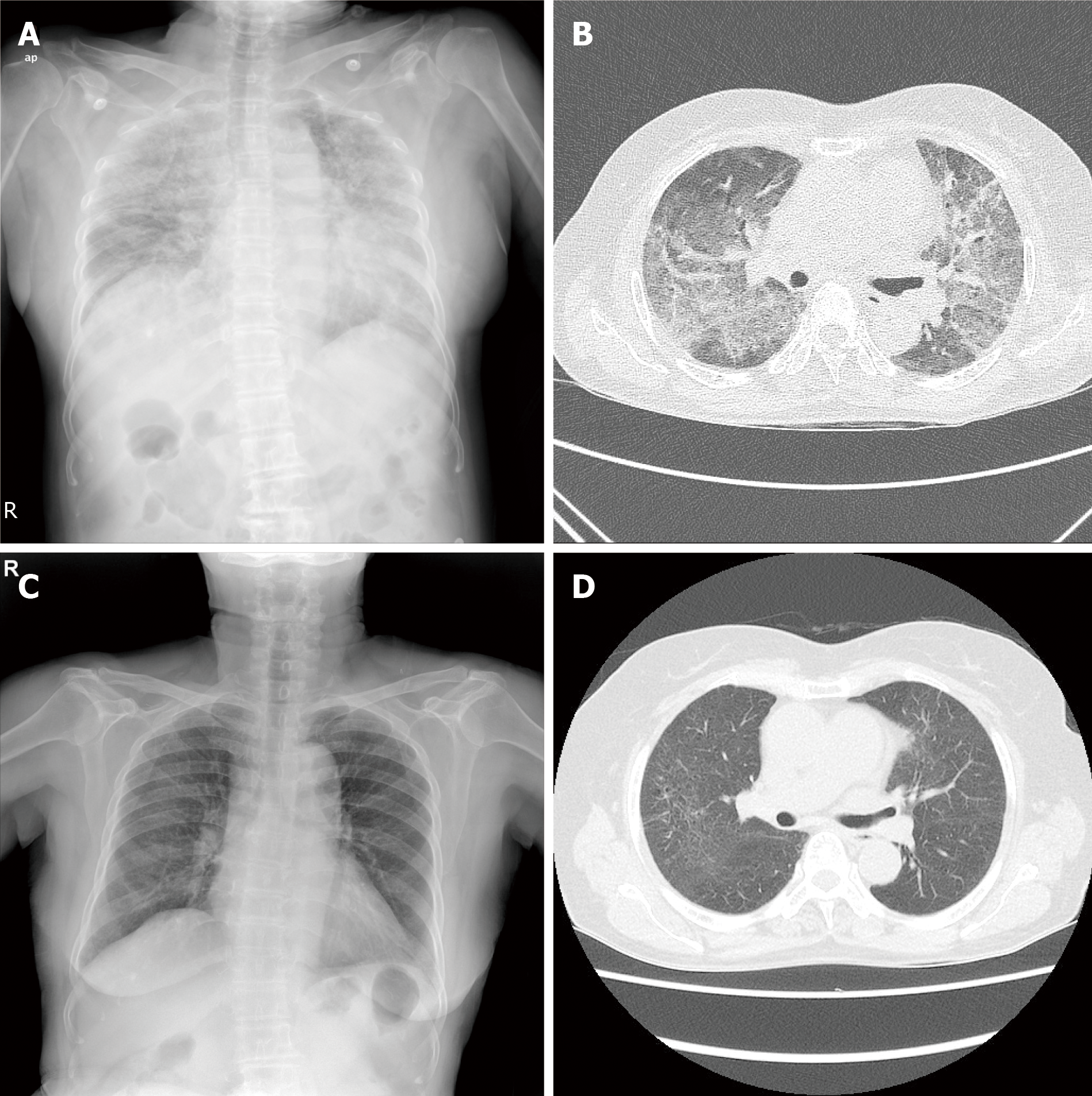
Figure 2 Chest X-ray and computed tomography images of Case 1.
A: At admission, chest radiography showed diffuse bilateral coalescent opacities; B: Contrast-enhanced chest computed tomography showed bilateral ground-glass opacities with interlobular interstitial thickening and patchy consolidation; C and D: After 4 mo, Chest radiography and high-resolution computed tomography showed almost improvement of ground-glass opacities, and patchy consolidation and peripheral reticulation were observed.
- Citation: Moon DS, Yoon SH, Lee SI, Park SG, Na YS. Interstitial lung disease induced by the roots of Achyranthes japonica Nakai: Three case reports. World J Clin Cases 2021; 9(8): 2015-2021
- URL: https://www.wjgnet.com/2307-8960/full/v9/i8/2015.htm
- DOI: https://dx.doi.org/10.12998/wjcc.v9.i8.2015


