Copyright
©The Author(s) 2021.
World J Clin Cases. Mar 16, 2021; 9(8): 2001-2007
Published online Mar 16, 2021. doi: 10.12998/wjcc.v9.i8.2001
Published online Mar 16, 2021. doi: 10.12998/wjcc.v9.i8.2001
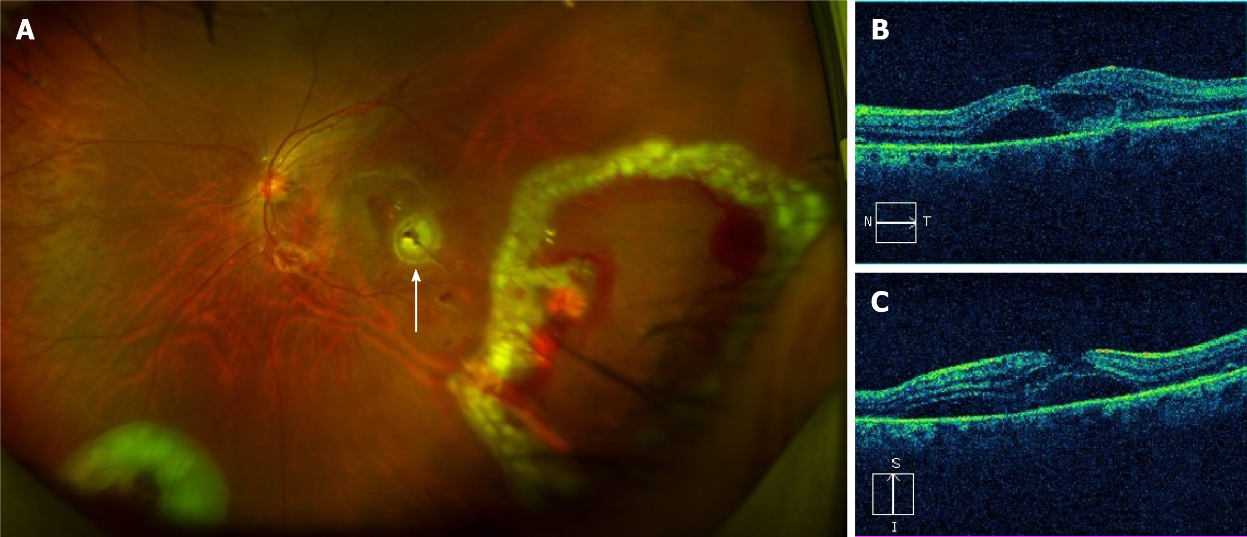
Figure 2 Fundus photography and optical coherence tomography obtained on the third postoperative day.
A: On the third postoperative day, fundus photography revealed the re-attached retina, the position of needle entry (white arrow), endo-laser spots, and minor hemorrhage; B and C: Optical coherence tomography revealed a small amount of fluid between the neuroepithelial layer of the retina and the retina pigment epithelium.
- Citation: Dai Y, Sun T, Gong JF. Inadvertent globe penetration during retrobulbar anesthesia: A case report . World J Clin Cases 2021; 9(8): 2001-2007
- URL: https://www.wjgnet.com/2307-8960/full/v9/i8/2001.htm
- DOI: https://dx.doi.org/10.12998/wjcc.v9.i8.2001


