Copyright
©The Author(s) 2021.
World J Clin Cases. Mar 16, 2021; 9(8): 1940-1945
Published online Mar 16, 2021. doi: 10.12998/wjcc.v9.i8.1940
Published online Mar 16, 2021. doi: 10.12998/wjcc.v9.i8.1940
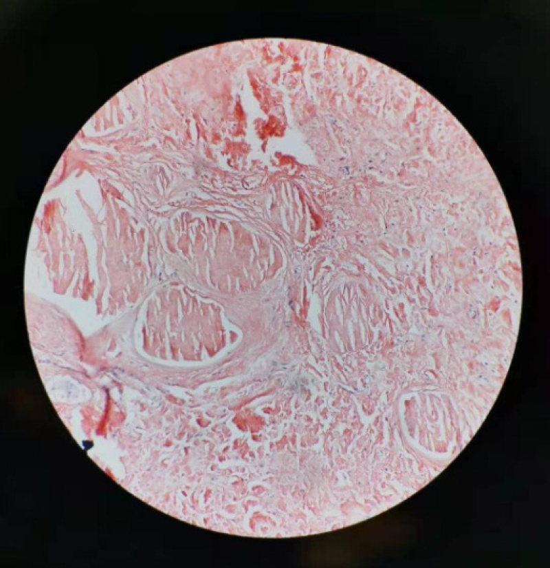
Figure 5 Photomicrograph (magnification, × 200) demonstrates homogeneous orange material without structure in coarse granular, massive, nodular forms on Congo red staining.
- Citation: Che ZG, Ni T, Wang ZC, Wang DW. Computed tomography imaging features for amyloid dacryolith in the nasolacrimal excretory system: A case report. World J Clin Cases 2021; 9(8): 1940-1945
- URL: https://www.wjgnet.com/2307-8960/full/v9/i8/1940.htm
- DOI: https://dx.doi.org/10.12998/wjcc.v9.i8.1940


