Copyright
©The Author(s) 2021.
World J Clin Cases. Mar 16, 2021; 9(8): 1931-1939
Published online Mar 16, 2021. doi: 10.12998/wjcc.v9.i8.1931
Published online Mar 16, 2021. doi: 10.12998/wjcc.v9.i8.1931
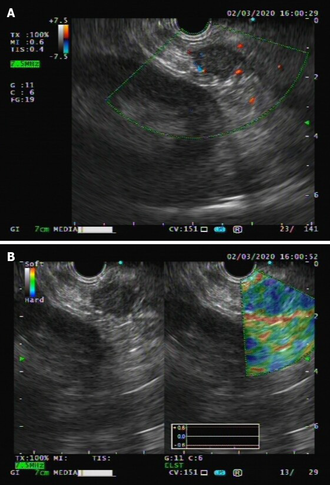
Figure 1 Endoscopic ultrasound images of the epithelioid angiomyolipoma.
A: Endoscopic ultrasound showed a hypoechogenic mass in the tail of the pancreas, measuring 1.9 cm, without obvious blood flow on color doppler flow imaging; B: Elastography showed that the elastic properties of the mass were a little harder than the surrounding tissue.
- Citation: Zhu QQ, Niu ZF, Yu FD, Wu Y, Wang GB. Epithelioid angiomyolipoma of the pancreas: A case report and review of the literature. World J Clin Cases 2021; 9(8): 1931-1939
- URL: https://www.wjgnet.com/2307-8960/full/v9/i8/1931.htm
- DOI: https://dx.doi.org/10.12998/wjcc.v9.i8.1931


