Copyright
©The Author(s) 2021.
World J Clin Cases. Mar 16, 2021; 9(8): 1885-1892
Published online Mar 16, 2021. doi: 10.12998/wjcc.v9.i8.1885
Published online Mar 16, 2021. doi: 10.12998/wjcc.v9.i8.1885
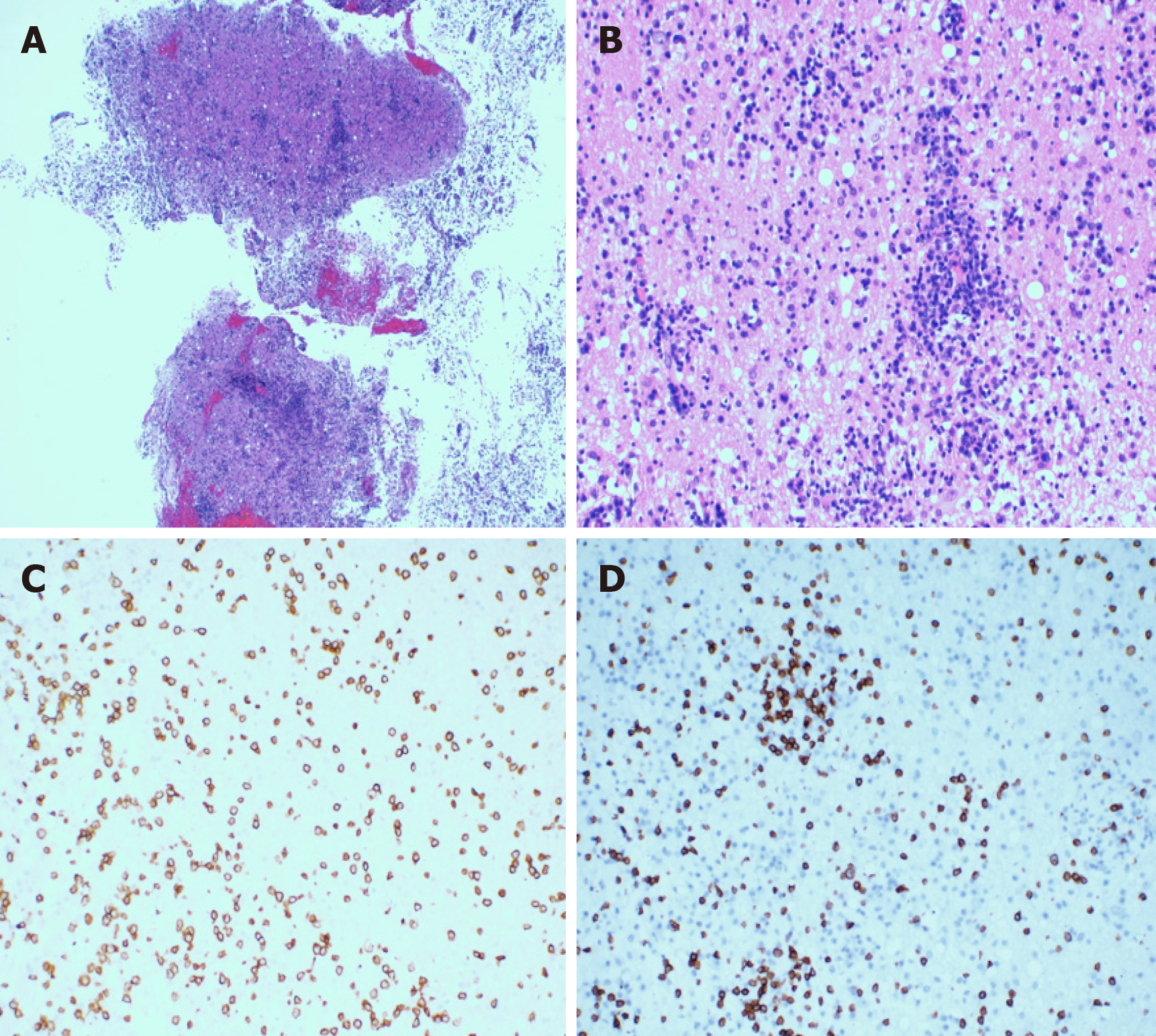
Figure 5 The hematoxylin-eosin staining of the resected specimens revealed that the plasmocytes and lymphocytes had proliferated.
A and B: The lymphocytes infiltrated the blood vessels and showed a vascular-centered growth pattern (A: 40 ×; B: 200 ×); C and D: Immunohistological staining showing the intracranial tumor positivity for CD3 and CD8 (original magnification 200 ×); C: CD3; D: CD8.
- Citation: Sun J, Ma XS, Qu LM, Song XS. Subcutaneous panniculitis-like T-cell lymphoma invading central nervous system in long-term clinical remission with lenalidomide: A case report. World J Clin Cases 2021; 9(8): 1885-1892
- URL: https://www.wjgnet.com/2307-8960/full/v9/i8/1885.htm
- DOI: https://dx.doi.org/10.12998/wjcc.v9.i8.1885


