Copyright
©The Author(s) 2021.
World J Clin Cases. Mar 16, 2021; 9(8): 1844-1852
Published online Mar 16, 2021. doi: 10.12998/wjcc.v9.i8.1844
Published online Mar 16, 2021. doi: 10.12998/wjcc.v9.i8.1844
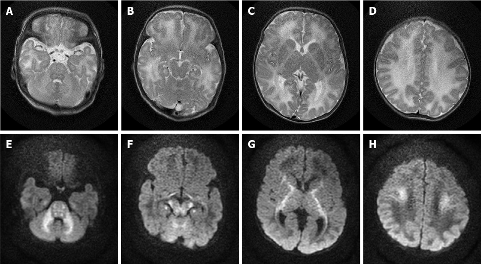
Figure 3 A 4-day-old male neonate with maple syrup urine disease.
Axial T2-weighted and axial diffusion-weighted MR images of the brain show bilateral symmetrical lesions involving the white matter of the cerebellum, pons, dorsal aspect of the mid brain, cerebral peduncles, corticospinal tract, internal capsule, and centrum semiovale. A-D: Axial T2-weighted magnetic resonance images; E-H: Axial diffusion-weighted magnetic resonance images.
- Citation: Li Y, Liu X, Duan CF, Song XF, Zhuang XH. Brain magnetic resonance imaging findings and radiologic review of maple syrup urine disease: Report of three cases. World J Clin Cases 2021; 9(8): 1844-1852
- URL: https://www.wjgnet.com/2307-8960/full/v9/i8/1844.htm
- DOI: https://dx.doi.org/10.12998/wjcc.v9.i8.1844


