Copyright
©The Author(s) 2021.
World J Clin Cases. Mar 16, 2021; 9(8): 1844-1852
Published online Mar 16, 2021. doi: 10.12998/wjcc.v9.i8.1844
Published online Mar 16, 2021. doi: 10.12998/wjcc.v9.i8.1844
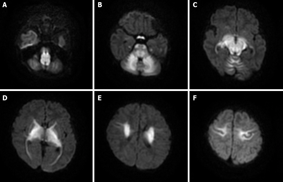
Figure 2 Axial diffusion-weighted sequences obtained in a 16-day-old female maple syrup urine disease patient.
Affected areas show abnormally increased signal intensity. A: Medulla; B: Cerebellar white matter, pons, and optic tracts; C: Midbrain; D-F: Internal capsule, thalami, globus palladi, and corona radiata.
- Citation: Li Y, Liu X, Duan CF, Song XF, Zhuang XH. Brain magnetic resonance imaging findings and radiologic review of maple syrup urine disease: Report of three cases. World J Clin Cases 2021; 9(8): 1844-1852
- URL: https://www.wjgnet.com/2307-8960/full/v9/i8/1844.htm
- DOI: https://dx.doi.org/10.12998/wjcc.v9.i8.1844


