Copyright
©The Author(s) 2021.
World J Clin Cases. Mar 16, 2021; 9(8): 1844-1852
Published online Mar 16, 2021. doi: 10.12998/wjcc.v9.i8.1844
Published online Mar 16, 2021. doi: 10.12998/wjcc.v9.i8.1844
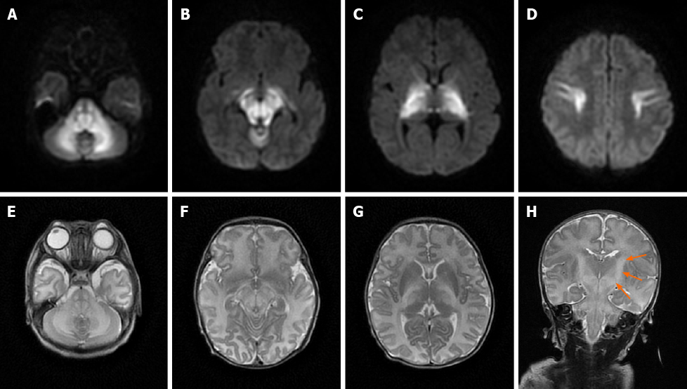
Figure 1 An 11-day-old male neonate with maple syrup urine disease.
Axial diffusion-weighted, axial T2-weighted, and coronal T2-weighted magnetic resonance images of the brain show bilateral symmetrical lesions involving the corticospinal tracts (arrows), thalami, globus pallidus, midbrain, dorsal brain stem, and cerebellar white matter. A-D: Axial diffusion-weighted magnetic resonance images; E-G: Axial T2-weighted magnetic resonance images; H: Coronal T2-weighted magnetic resonance images.
- Citation: Li Y, Liu X, Duan CF, Song XF, Zhuang XH. Brain magnetic resonance imaging findings and radiologic review of maple syrup urine disease: Report of three cases. World J Clin Cases 2021; 9(8): 1844-1852
- URL: https://www.wjgnet.com/2307-8960/full/v9/i8/1844.htm
- DOI: https://dx.doi.org/10.12998/wjcc.v9.i8.1844


