Copyright
©The Author(s) 2021.
World J Clin Cases. Mar 6, 2021; 9(7): 1734-1740
Published online Mar 6, 2021. doi: 10.12998/wjcc.v9.i7.1734
Published online Mar 6, 2021. doi: 10.12998/wjcc.v9.i7.1734
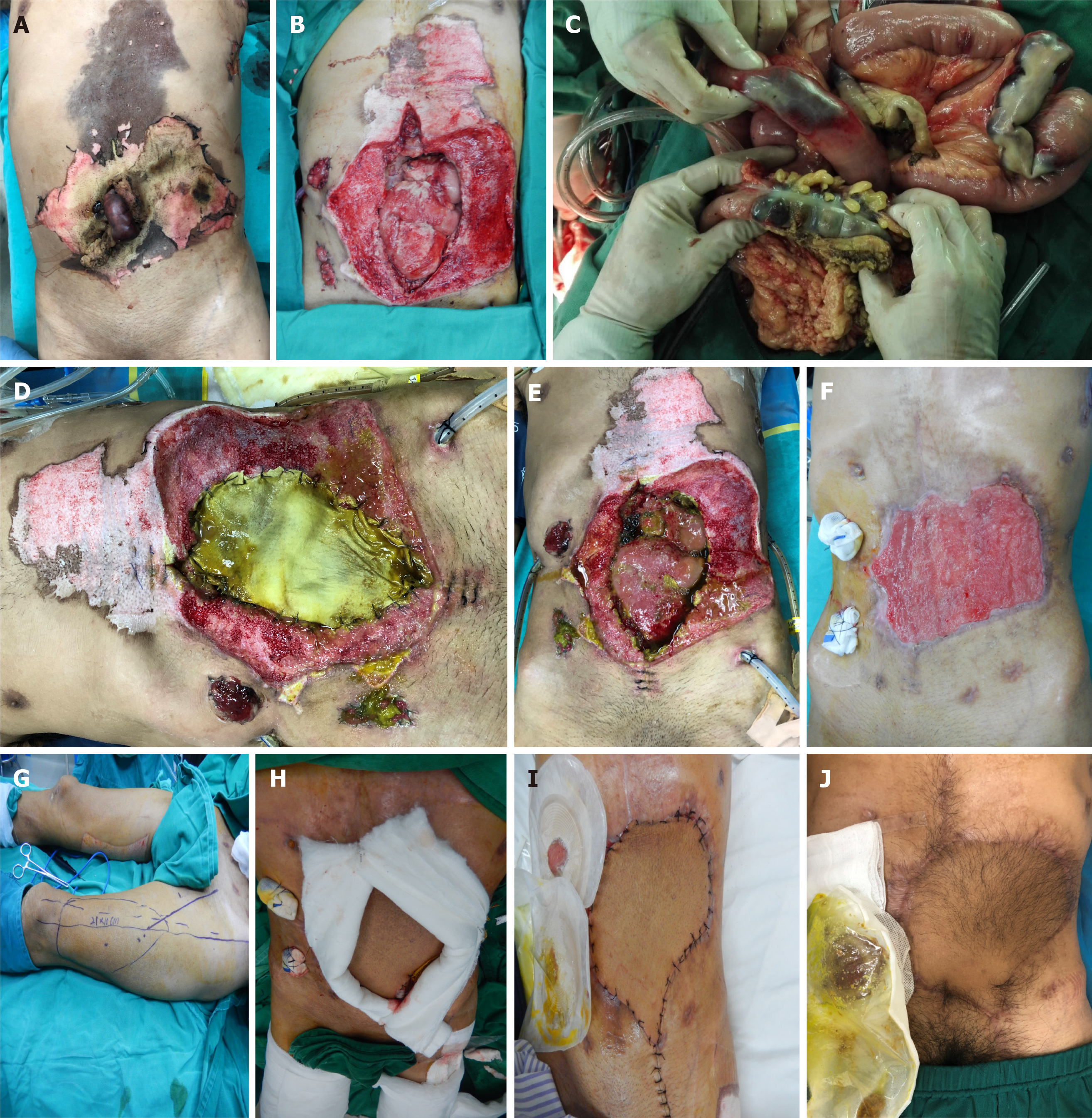
Figure 1 Operation situation.
A: Necrotic bowels exposed through the ruptured eschar of the electrically burned abdominal wall; B: Huge abdominal wall defects and exposed bowels following debridement; C: Necrosis of multiple bowel segments revealed during the exploratory laparotomy; D: Acellular dermal matrix (ADM) contaminated with the duodenal leaks 5 d after the surgery; E: Reduced duodenal leak and granulation tissue growth on the serosal surfaces of part of the bowels after treatments such as ADM closure of the bowels, vacuum sealing drainage irrigation, and abdominal drainage 4 wk after the surgery; F: Dense granulation tissues on the bowel serosal surface, forming a plate-like adhesive barrier that completely enclosed the abdominal cavity 6 wk after the surgery; G: Design of the left-side anterolateral thigh (ALT) flap; H: Repair of abdominal wall defects using a combined ALT and tensor fasciae lata free flap; I: Flap survived well 1 wk after the surgery; J: Excellent repair and reconstruction of the abdominal wall without abdominal hernia or bowel obstruction at the 6 mo follow-up.
- Citation: Wang J. Reconstructing abdominal wall defects with a free composite tissue flap: A case report. World J Clin Cases 2021; 9(7): 1734-1740
- URL: https://www.wjgnet.com/2307-8960/full/v9/i7/1734.htm
- DOI: https://dx.doi.org/10.12998/wjcc.v9.i7.1734


