Copyright
©The Author(s) 2021.
World J Clin Cases. Feb 26, 2021; 9(6): 1439-1445
Published online Feb 26, 2021. doi: 10.12998/wjcc.v9.i6.1439
Published online Feb 26, 2021. doi: 10.12998/wjcc.v9.i6.1439
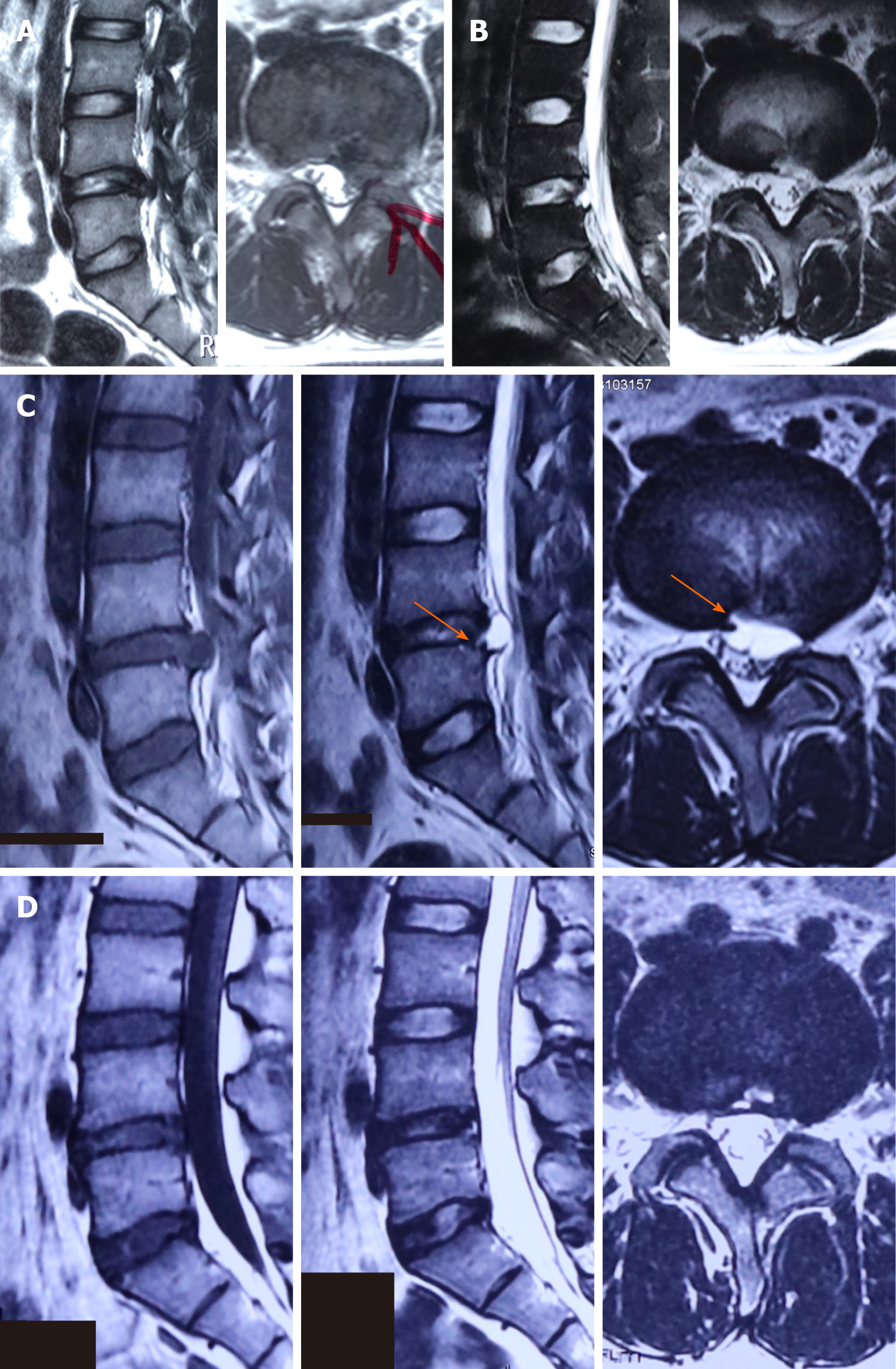
Figure 1 Magnetic resonance imaging of case 1.
A: Pre-surgical magnetic resonance imaging (MRI) showing lumbar disc herniation at L4-5; B: Post-surgical MRI confirming complete removal of the herniated disc fragment; C: Re-examination MRI revealed a cystic lesion at the surgical site with a communication stalk between the cyst and inner disc (arrow); D: Follow-up MRI after 6 mo demonstrating complete spontaneous regression of the lesion.
- Citation: Fu CF, Tian ZS, Yao LY, Yao JH, Jin YZ, Liu Y, Wang YY. Postoperative discal pseudocyst and its similarities to discal cyst: A case report . World J Clin Cases 2021; 9(6): 1439-1445
- URL: https://www.wjgnet.com/2307-8960/full/v9/i6/1439.htm
- DOI: https://dx.doi.org/10.12998/wjcc.v9.i6.1439


