Copyright
©The Author(s) 2021.
World J Clin Cases. Feb 26, 2021; 9(6): 1433-1438
Published online Feb 26, 2021. doi: 10.12998/wjcc.v9.i6.1433
Published online Feb 26, 2021. doi: 10.12998/wjcc.v9.i6.1433
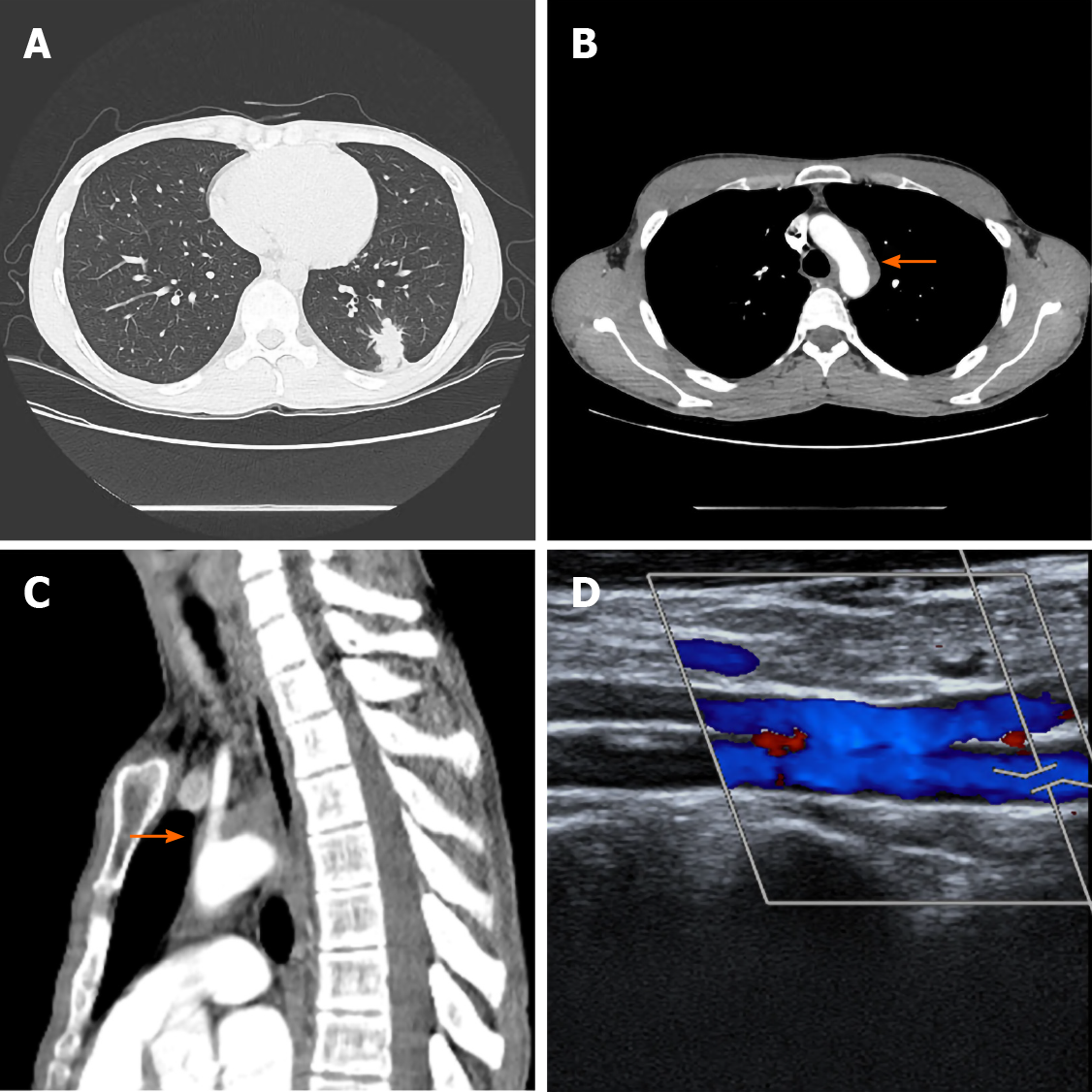
Figure 1 Initial computed tomography scans.
A: Chest computed tomography scan with contrast enhancement showing a nodule abutting the pleura in the left lower lobe of the lung; B and C: Computed tomography scan showing a heterogeneously enhancing soft tissue dense lesion partially occluding the aortic arch; and D: Doppler ultrasonography for the evaluation of subclavian steal syndrome demonstrated a reverse flow in the left vertebral artery.
- Citation: Cho U, Kim SK, Ko JM, Yoo J. Unusual presentation of granulomatosis with polyangiitis causing periaortitis and consequent subclavian steal syndrome: A case report. World J Clin Cases 2021; 9(6): 1433-1438
- URL: https://www.wjgnet.com/2307-8960/full/v9/i6/1433.htm
- DOI: https://dx.doi.org/10.12998/wjcc.v9.i6.1433


