Copyright
©The Author(s) 2021.
World J Clin Cases. Feb 26, 2021; 9(6): 1424-1432
Published online Feb 26, 2021. doi: 10.12998/wjcc.v9.i6.1424
Published online Feb 26, 2021. doi: 10.12998/wjcc.v9.i6.1424
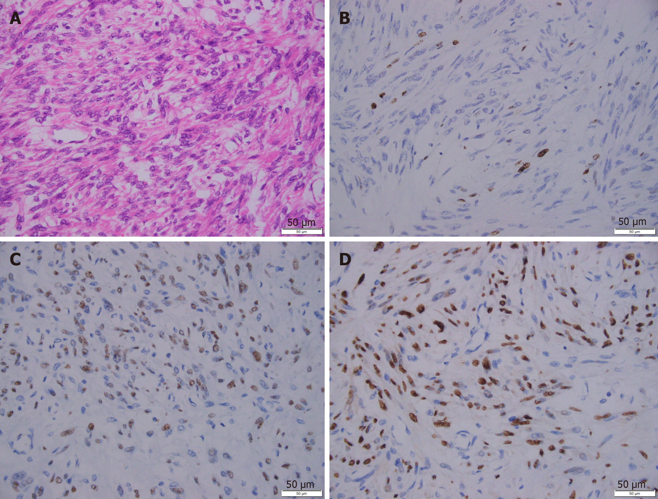
Figure 4 Representative immunohistochemical images of the leiomyoma.
A: H&E staining; B: Ki67 staining; C: Estrogen receptor staining; D: Progesterone receptor staining. Scale bar: 50 μm.
- Citation: Wang YW, Fan Q, Qian ZX, Wang JJ, Li YH, Wang YD. Abdominopelvic leiomyoma with large ascites: A case report and review of the literature. World J Clin Cases 2021; 9(6): 1424-1432
- URL: https://www.wjgnet.com/2307-8960/full/v9/i6/1424.htm
- DOI: https://dx.doi.org/10.12998/wjcc.v9.i6.1424


