Copyright
©The Author(s) 2021.
World J Clin Cases. Feb 26, 2021; 9(6): 1336-1342
Published online Feb 26, 2021. doi: 10.12998/wjcc.v9.i6.1336
Published online Feb 26, 2021. doi: 10.12998/wjcc.v9.i6.1336
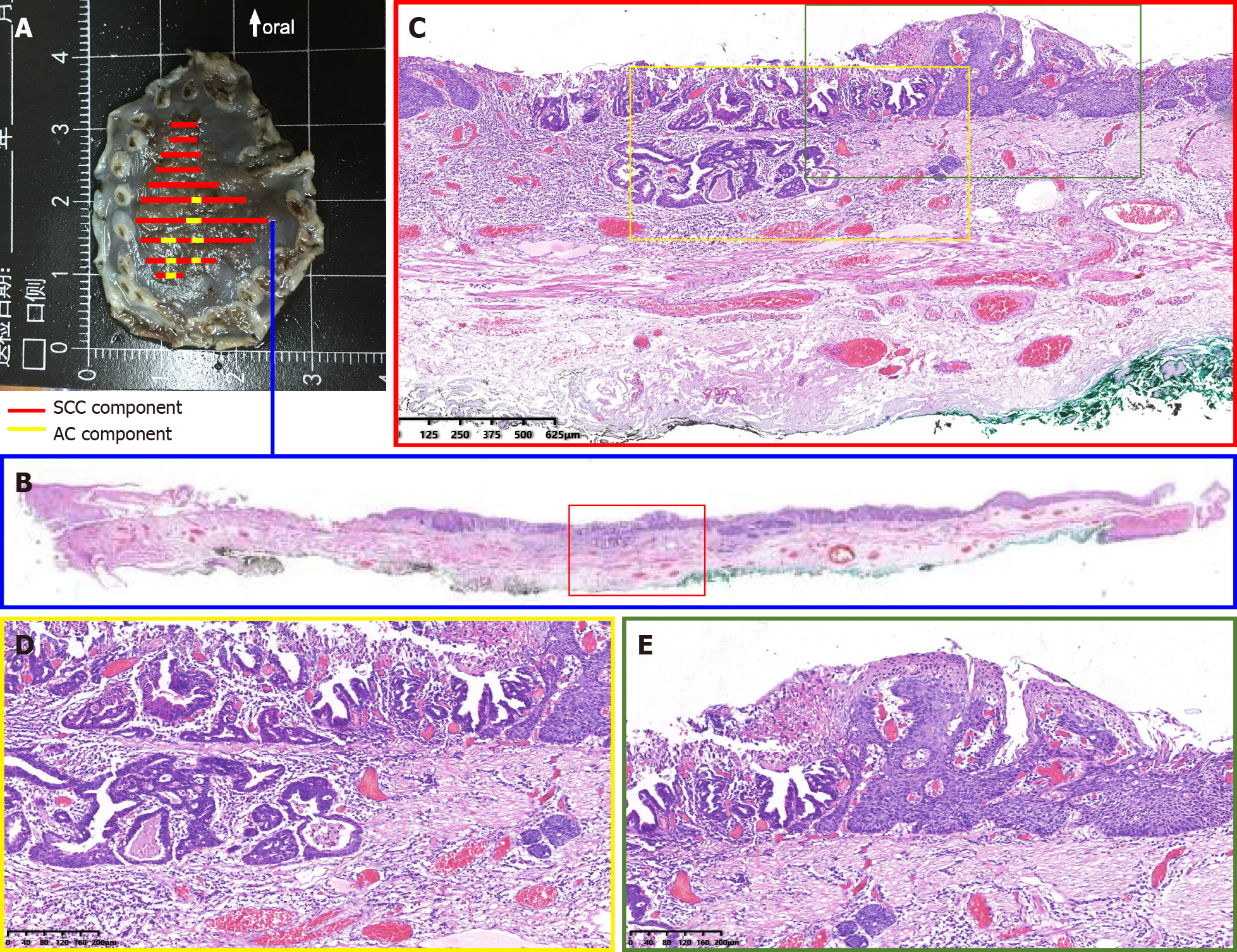
Figure 3 Dilated and tortuous vessels were observed around and within the ductal-structure-forming cancer cell nests.
A: Specimen resected by endoscopic submucosal dissection; B: Histological view of the resected lesion revealed that the squamous cell carcinoma element was the predominant element, and the adenocarcinoma element was distributed in a focal pattern among the squamous cell carcinoma element; C-E: High-power view revealed invasive carcinoma forming ductal structures distributed among squamous cell carcinoma. Part of the lesion was covered by dyskeratotic squamous cells.
- Citation: Liu GY, Zhang JX, Rong L, Nian WD, Nian BX, Tian Y. Esophageal superficial adenosquamous carcinoma resected by endoscopic submucosal dissection: A rare case report. World J Clin Cases 2021; 9(6): 1336-1342
- URL: https://www.wjgnet.com/2307-8960/full/v9/i6/1336.htm
- DOI: https://dx.doi.org/10.12998/wjcc.v9.i6.1336


