Copyright
©The Author(s) 2021.
World J Clin Cases. Feb 26, 2021; 9(6): 1336-1342
Published online Feb 26, 2021. doi: 10.12998/wjcc.v9.i6.1336
Published online Feb 26, 2021. doi: 10.12998/wjcc.v9.i6.1336
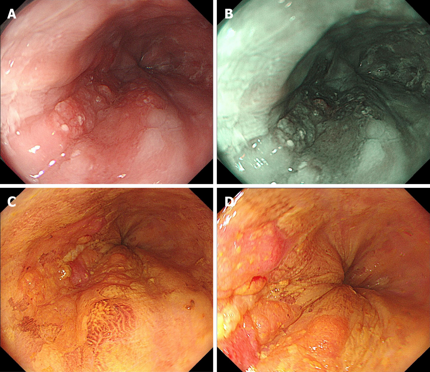
Figure 1 Esophagogastroduodenoscopy revealing a superficial lesion in the distal esophagus.
A: Upon white light endoscopy, a flat lesion of approximately 2.5 cm × 1.5 cm in size was found, with the central part uneven and slightly elevated. The lesion presented slight reddening of the mucosa with a coarse surface; B: Narrow-band imaging showed a well-demarcated brownish area of background coloration; C and D: Iodine staining (1%) visualized the lesion as an unstained area with a comparatively clear boundary, and pink color sign could be seen.
- Citation: Liu GY, Zhang JX, Rong L, Nian WD, Nian BX, Tian Y. Esophageal superficial adenosquamous carcinoma resected by endoscopic submucosal dissection: A rare case report. World J Clin Cases 2021; 9(6): 1336-1342
- URL: https://www.wjgnet.com/2307-8960/full/v9/i6/1336.htm
- DOI: https://dx.doi.org/10.12998/wjcc.v9.i6.1336


