Copyright
©The Author(s) 2021.
World J Clin Cases. Feb 16, 2021; 9(5): 1184-1195
Published online Feb 16, 2021. doi: 10.12998/wjcc.v9.i5.1184
Published online Feb 16, 2021. doi: 10.12998/wjcc.v9.i5.1184
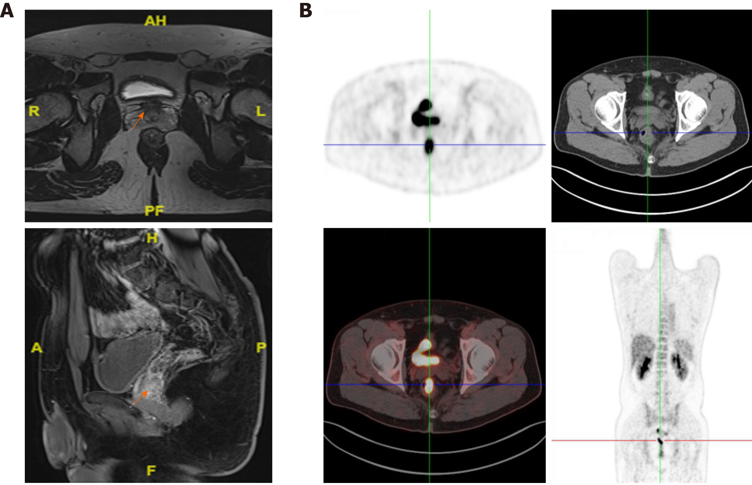
Figure 3 Axial computed tomography images reveal a rectal mass.
A: Axial maximum intensive projection at the same imaging level, and coronal images showed a 40 mm solitary nodule approximately 58 mm away above the anus in the rectum; B: Axial images of positron emission topography-computed tomography at the same imaging level, and rectal image of positron emission topography-computed tomography maximum intensive projection showed a thickened medial rectum wall with homogeneous uptake of 18F-fluorodeoxy-glucose, and the maximum standardized uptake value was 14.6.
- Citation: Peng WX, Liu X, Wang QF, Zhou XY, Luo ZG, Hu XC. Heterochronic triple primary malignancies with Epstein-Barr virus infection and tumor protein 53 gene mutation: A case report and review of literature. World J Clin Cases 2021; 9(5): 1184-1195
- URL: https://www.wjgnet.com/2307-8960/full/v9/i5/1184.htm
- DOI: https://dx.doi.org/10.12998/wjcc.v9.i5.1184


