Copyright
©The Author(s) 2021.
World J Clin Cases. Feb 16, 2021; 9(5): 1168-1174
Published online Feb 16, 2021. doi: 10.12998/wjcc.v9.i5.1168
Published online Feb 16, 2021. doi: 10.12998/wjcc.v9.i5.1168
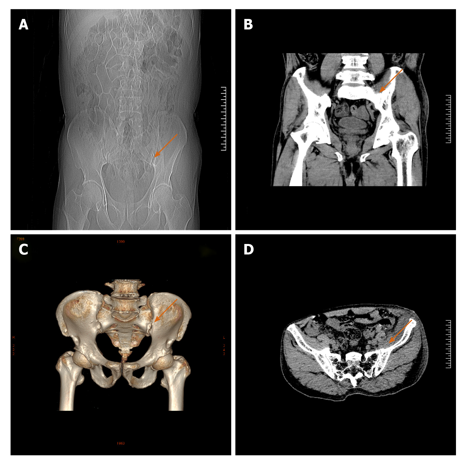
Figure 5 Pelvic computed tomography scan plus 3D reconstruction after the operation.
A: Anteroposterior radiograph of the pelvis; B: Coronal computed tomography scan showed the osteophyte has been removed; C: 3D reconstruction showed that the osteophyte from the anterior lower part of the left sacroiliac joint has been completely removed and the articular surface was flat while the osteophyte of the right was still existing, as indicated by the orange arrow; D: Horizontal scan of the pelvic after surgery.
- Citation: Cai MD, Zhang HF, Fan YG, Su XJ, Xia L. Obturator nerve impingement caused by an osteophyte in the sacroiliac joint: A case report. World J Clin Cases 2021; 9(5): 1168-1174
- URL: https://www.wjgnet.com/2307-8960/full/v9/i5/1168.htm
- DOI: https://dx.doi.org/10.12998/wjcc.v9.i5.1168


