Copyright
©The Author(s) 2021.
World J Clin Cases. Feb 16, 2021; 9(5): 1132-1138
Published online Feb 16, 2021. doi: 10.12998/wjcc.v9.i5.1132
Published online Feb 16, 2021. doi: 10.12998/wjcc.v9.i5.1132
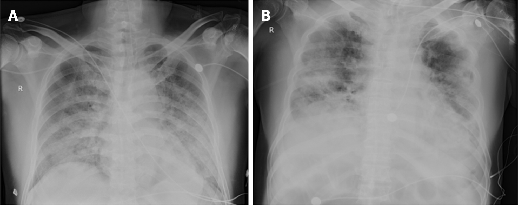
Figure 1 Chest imaging.
A: Bedside chest image of the patient, taken on February 3, 2020, i.e., the 13th day of the disease, showing multiple patchy ground-glass opacities in the bilateral lungs, and some of them were with consolidation; B: Chest image of the patient, taken on February 28, 2020, i.e., the 38th day of the disease, showing multiple exudative lesions in bilateral lungs and local consolidations, and the lesions in lateral strips of bilateral lungs increased.
- Citation: Chen QY, He YS, Liu K, Cao J, Chen YX. Bronchoscopy for diagnosis of COVID-19 with respiratory failure: A case report. World J Clin Cases 2021; 9(5): 1132-1138
- URL: https://www.wjgnet.com/2307-8960/full/v9/i5/1132.htm
- DOI: https://dx.doi.org/10.12998/wjcc.v9.i5.1132


