Copyright
©The Author(s) 2021.
World J Clin Cases. Feb 16, 2021; 9(5): 1111-1118
Published online Feb 16, 2021. doi: 10.12998/wjcc.v9.i5.1111
Published online Feb 16, 2021. doi: 10.12998/wjcc.v9.i5.1111
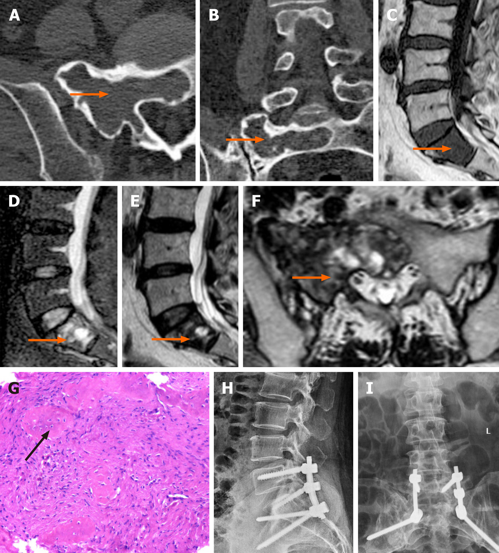
Figure 1 A 60-year-old female with monostotic fibrous dysplasia in the sacrum.
A and B: Computed tomography images showing an expansile lesion with a sclerotic rim and ground glass opacity; C: Sagittal T1WI showing low signal intensity; D-F: Sagittal T2WI, FS T2WI and axial T2WI showing iso-high signal intensity; G: Pathological haematoxylin-eosin (HE) staining (×10) showing tumour-like hyperplasia of fibrous tissue with surrounding fibrotic bone formation (arrow); H and I: After the operation, the patient underwent an X-ray examination.
- Citation: Liu XX, Xin X, Yan YH, Ma XW. Imaging characteristics of a rare case of monostotic fibrous dysplasia of the sacrum: A case report. World J Clin Cases 2021; 9(5): 1111-1118
- URL: https://www.wjgnet.com/2307-8960/full/v9/i5/1111.htm
- DOI: https://dx.doi.org/10.12998/wjcc.v9.i5.1111


