Copyright
©The Author(s) 2021.
World J Clin Cases. Feb 16, 2021; 9(5): 1079-1086
Published online Feb 16, 2021. doi: 10.12998/wjcc.v9.i5.1079
Published online Feb 16, 2021. doi: 10.12998/wjcc.v9.i5.1079
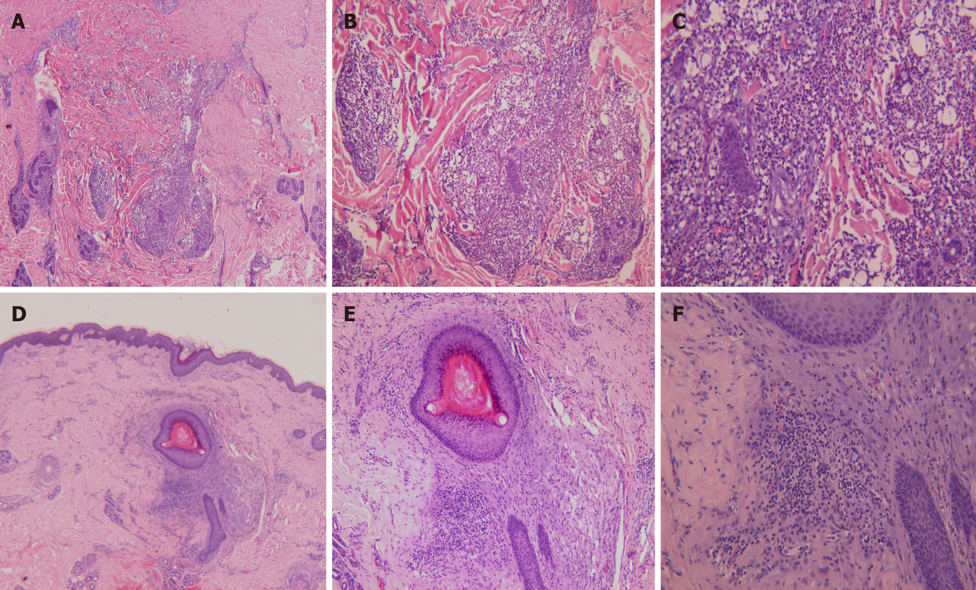
Figure 3 Histopathological staining of the skin eruption.
A-C: Histopathological images of the back skin eruption revealing hyperkeratinization, follicular keratotic plugs, focal epidermal hyperproliferation, vasodilatation and hyperemia in the dermis, destruction of hair follicles and sebaceous glands, and massive perifollicular neutrophil-dominant inflammatory infiltration, suggesting folliculitis and perifolliculitis (A, hematoxylin-eosin [HE] × 40; B, HE × 100; C, HE × 200); D-F: Histopathological images of the ear skin eruption revealing fairly normal epidermis and perifollicular lymphocyte-dominant inflammatory infiltration, suggesting perifolliculitis (D, HE × 40; E, HE × 100; F, HE × 200).
- Citation: Ma Y, Cao X, Zhang L, Zhang JY, Qiao ZS, Feng WL. Neuropathy and chloracne induced by 3,5,6-trichloropyridin-2-ol sodium: Report of three cases. World J Clin Cases 2021; 9(5): 1079-1086
- URL: https://www.wjgnet.com/2307-8960/full/v9/i5/1079.htm
- DOI: https://dx.doi.org/10.12998/wjcc.v9.i5.1079


