Copyright
©The Author(s) 2021.
World J Clin Cases. Feb 16, 2021; 9(5): 1037-1047
Published online Feb 16, 2021. doi: 10.12998/wjcc.v9.i5.1037
Published online Feb 16, 2021. doi: 10.12998/wjcc.v9.i5.1037
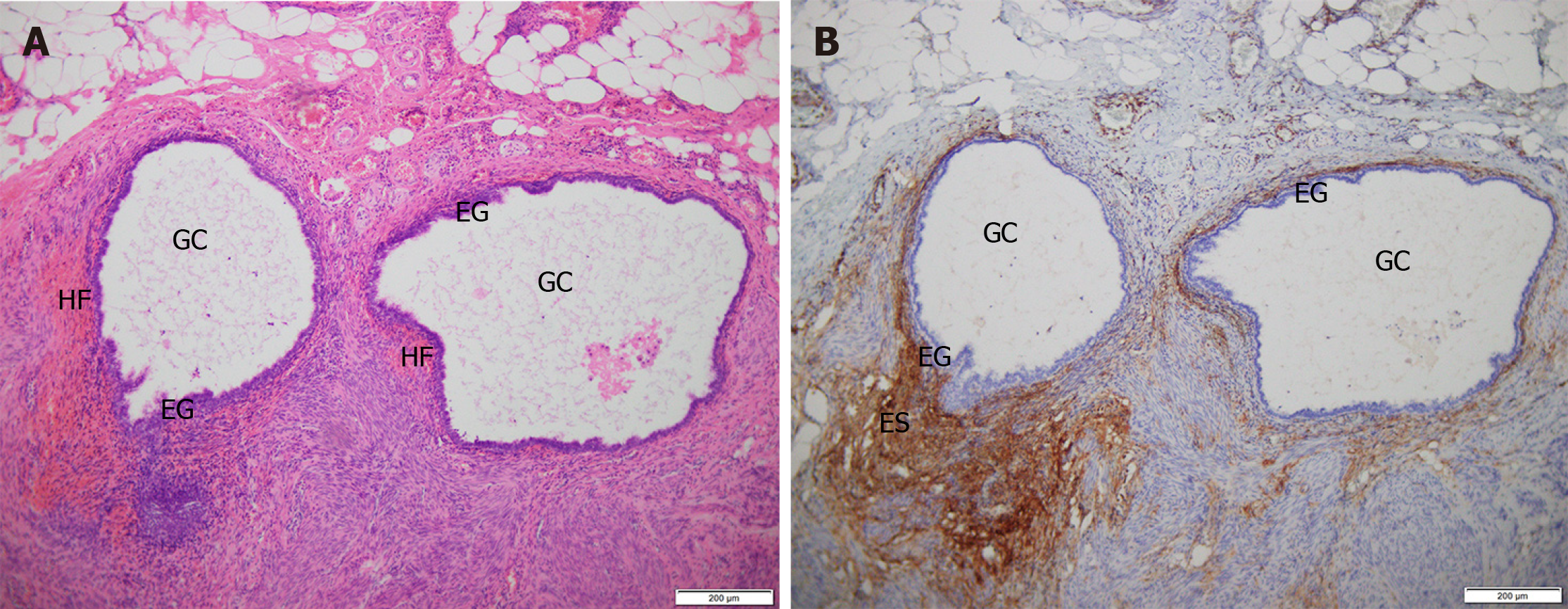
Figure 4 Hematoxylin-eosin staining and immunohistochemistry staining of perineal endometriosis lesions.
A: Hematoxylin-eosin (HE) staining shows endometriotic lesions are located in fat, smooth muscle and fibrous connective tissue. The lesion is composed of endometrial glands and surrounding stroma with local hemorrhage (HE, × 100); B: Immunohistochemistry staining shows that endometrial stromal cells are CD10 positive (× 100). GC: Glandular cavity; EG: Endometrial glands; ES: Endometrial stroma; HF: Hemorrhagic foci.
- Citation: Liang Y, Zhang D, Jiang L, Liu Y, Zhang J. Clinical characteristics of perineal endometriosis: A case series. World J Clin Cases 2021; 9(5): 1037-1047
- URL: https://www.wjgnet.com/2307-8960/full/v9/i5/1037.htm
- DOI: https://dx.doi.org/10.12998/wjcc.v9.i5.1037


