Copyright
©The Author(s) 2021.
World J Clin Cases. Feb 16, 2021; 9(5): 1037-1047
Published online Feb 16, 2021. doi: 10.12998/wjcc.v9.i5.1037
Published online Feb 16, 2021. doi: 10.12998/wjcc.v9.i5.1037
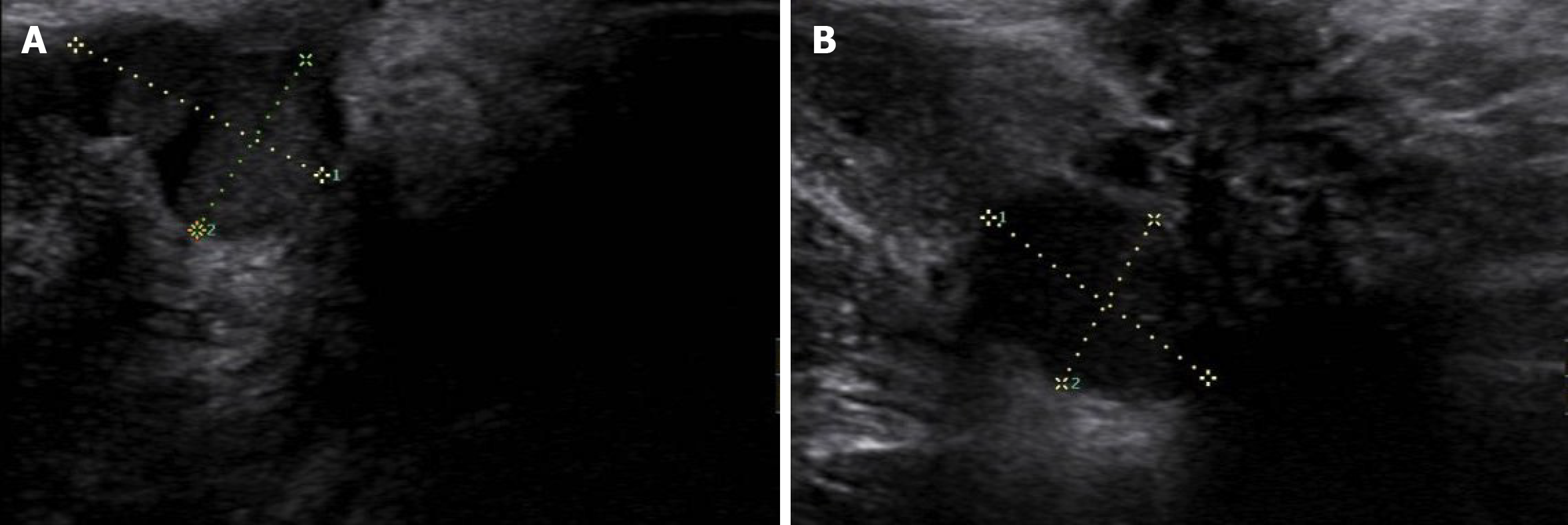
Figure 2 Ultrasound images of perineal endometriosis.
A: A 16 mm × 16 mm × 12 mm low-echo mass located under the perineal incision; B: Another heterogeneous-echo mass with an obscure boundary and regular shape is located beneath the lesion shown in Figure A.
- Citation: Liang Y, Zhang D, Jiang L, Liu Y, Zhang J. Clinical characteristics of perineal endometriosis: A case series. World J Clin Cases 2021; 9(5): 1037-1047
- URL: https://www.wjgnet.com/2307-8960/full/v9/i5/1037.htm
- DOI: https://dx.doi.org/10.12998/wjcc.v9.i5.1037


