Copyright
©The Author(s) 2021.
World J Clin Cases. Feb 6, 2021; 9(4): 992-998
Published online Feb 6, 2021. doi: 10.12998/wjcc.v9.i4.992
Published online Feb 6, 2021. doi: 10.12998/wjcc.v9.i4.992
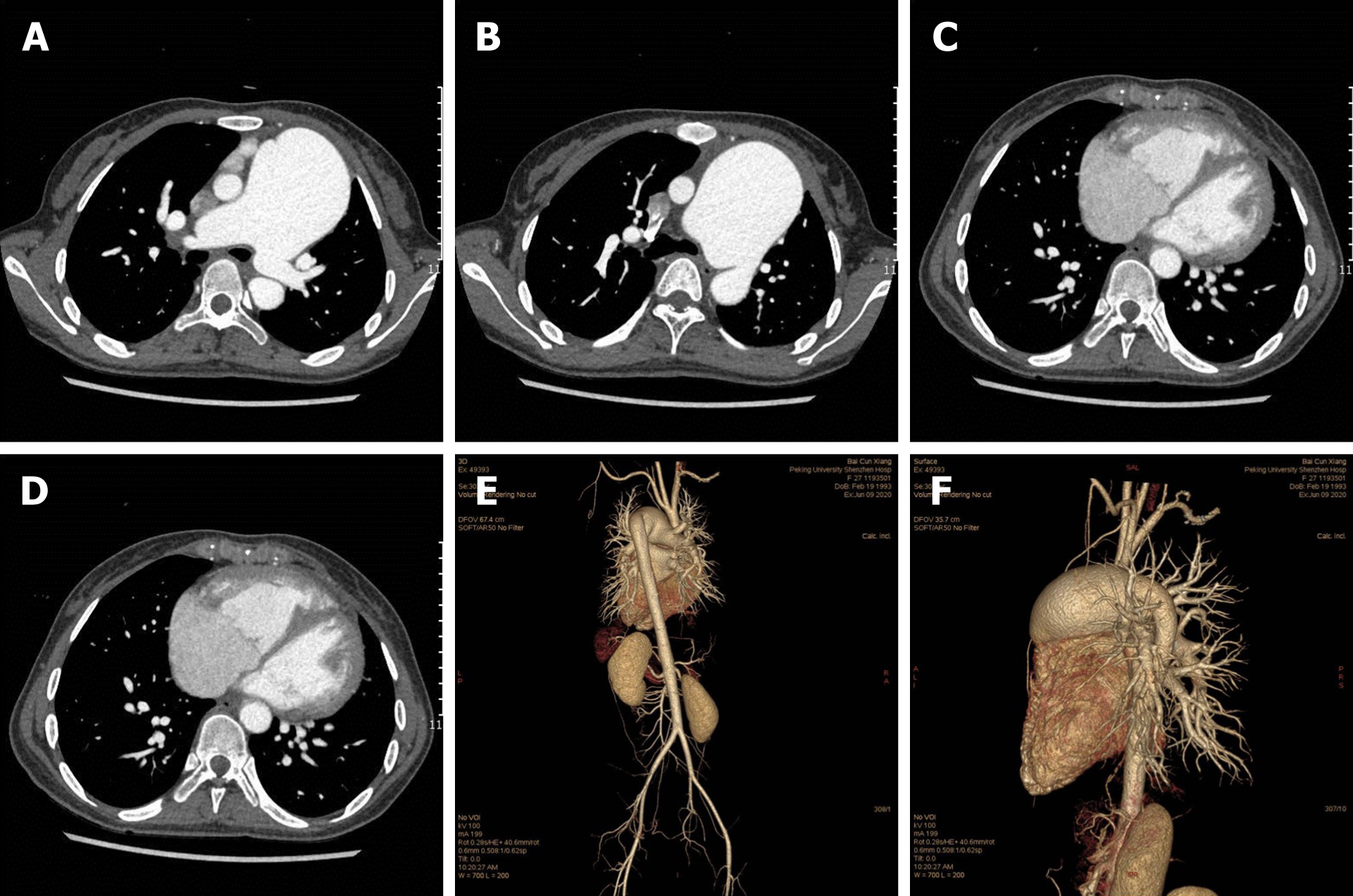
Figure 3 Computed tomography angiography.
A: The diameter of the main pulmonary artery and the right pulmonary artery are thickened at the level of the pulmonary trunk bifurcation; B: The descending aorta originates from the pulmonary trunk; C: The ventricular septal defect is seen at the four-chamber level of the heart; D: Axial image shows enlargement of the right heart; E: Volume-rendered image from a back projection depicts the interruption of the aortic arch and the descending aorta arising from the pulmonary trunk; F: Volume-rendered image from a left lateral projection depicts the pulmonary artery straddle.
- Citation: Dong SW, Di DD, Cheng GX. Isolated interrupted aortic arch in an adult: A case report. World J Clin Cases 2021; 9(4): 992-998
- URL: https://www.wjgnet.com/2307-8960/full/v9/i4/992.htm
- DOI: https://dx.doi.org/10.12998/wjcc.v9.i4.992


