Copyright
©The Author(s) 2021.
World J Clin Cases. Feb 6, 2021; 9(4): 992-998
Published online Feb 6, 2021. doi: 10.12998/wjcc.v9.i4.992
Published online Feb 6, 2021. doi: 10.12998/wjcc.v9.i4.992
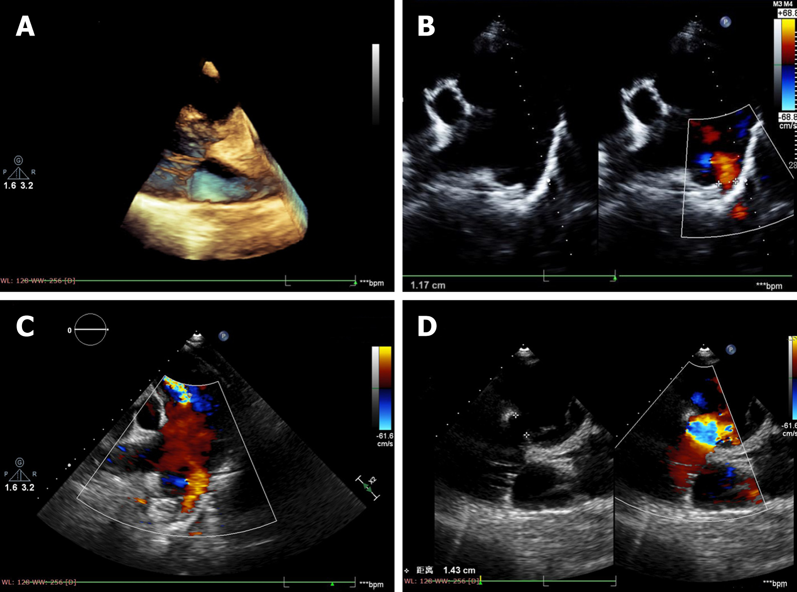
Figure 1 Three-dimensional ultrasound reconstruction.
A: The interrupted aortic arch; B: Color Doppler flow imaging shows an abnormal passage between the descending aorta and pulmonary artery; C: The main pulmonary artery and its branches are dilated at the level of bifurcation of the pulmonary trunk; D: The continuity of the ventricular septal outflow tract is interrupted and the defect size is about 14 mm.
- Citation: Dong SW, Di DD, Cheng GX. Isolated interrupted aortic arch in an adult: A case report. World J Clin Cases 2021; 9(4): 992-998
- URL: https://www.wjgnet.com/2307-8960/full/v9/i4/992.htm
- DOI: https://dx.doi.org/10.12998/wjcc.v9.i4.992


