Copyright
©The Author(s) 2021.
World J Clin Cases. Feb 6, 2021; 9(4): 927-934
Published online Feb 6, 2021. doi: 10.12998/wjcc.v9.i4.927
Published online Feb 6, 2021. doi: 10.12998/wjcc.v9.i4.927
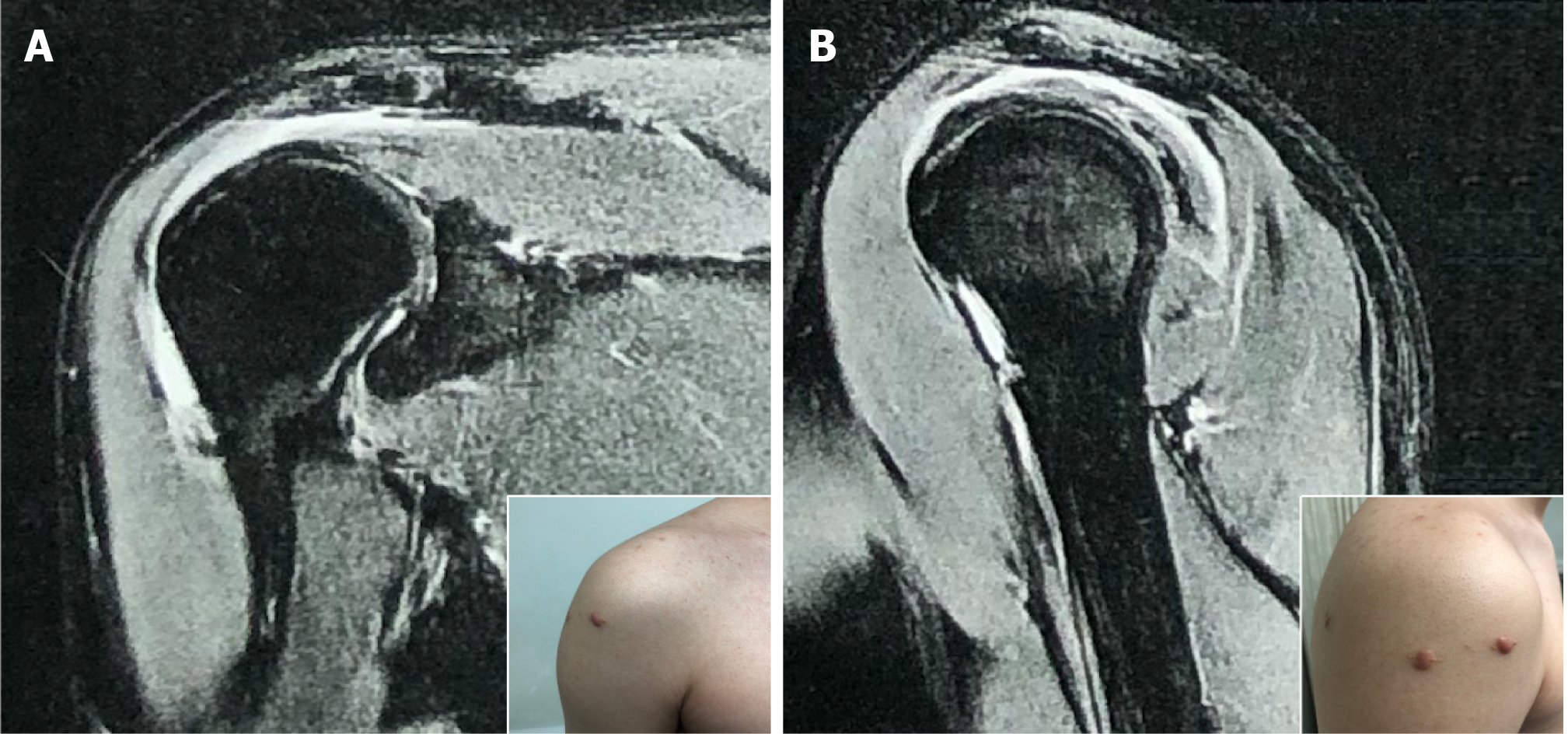
Figure 4 Follow-up imaging evaluation at 4 mo postoperative.
Magnetic resonance imaging showed the effusion of the subacromial bursa to be significantly reduced. A: Frontal view; B: Sagittal view. Corresponding insets show the incisions to have healed well after surgery.
- Citation: Wang FS, Shahzad K, Zhang WG, Li J, Tian K. Atypical presentation of shoulder brucellosis misdiagnosed as subacromial bursitis: A case report . World J Clin Cases 2021; 9(4): 927-934
- URL: https://www.wjgnet.com/2307-8960/full/v9/i4/927.htm
- DOI: https://dx.doi.org/10.12998/wjcc.v9.i4.927


