Copyright
©The Author(s) 2021.
World J Clin Cases. Feb 6, 2021; 9(4): 912-918
Published online Feb 6, 2021. doi: 10.12998/wjcc.v9.i4.912
Published online Feb 6, 2021. doi: 10.12998/wjcc.v9.i4.912
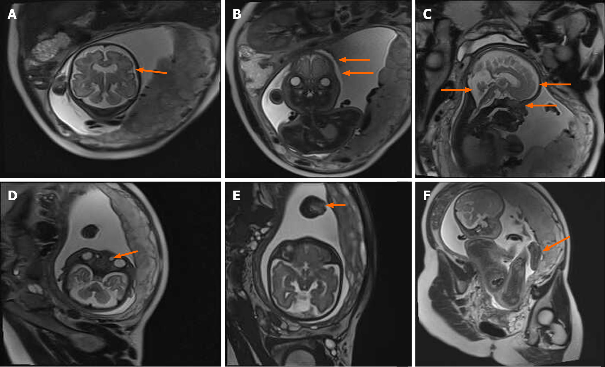
Figure 2 Magnetic resonance imaging performed at 31, 1/7 wk of pregnancy showed the following: A: Acrocephalia; B: Craniosynostosis, widened eye distances; C: Protruding forehead, concave nasal root, widened anterior horn of the lateral ventricle and the posterior cranial fossa; D: Nasolacrimal duct cyst; E: Syndactyly of hands; and F: Syndactyly of feet.
- Citation: Chen L, Huang FX. Apert syndrome diagnosed by prenatal ultrasound combined with magnetic resonance imaging and whole exome sequencing: A case report. World J Clin Cases 2021; 9(4): 912-918
- URL: https://www.wjgnet.com/2307-8960/full/v9/i4/912.htm
- DOI: https://dx.doi.org/10.12998/wjcc.v9.i4.912


