Copyright
©The Author(s) 2021.
World J Clin Cases. Feb 6, 2021; 9(4): 871-877
Published online Feb 6, 2021. doi: 10.12998/wjcc.v9.i4.871
Published online Feb 6, 2021. doi: 10.12998/wjcc.v9.i4.871
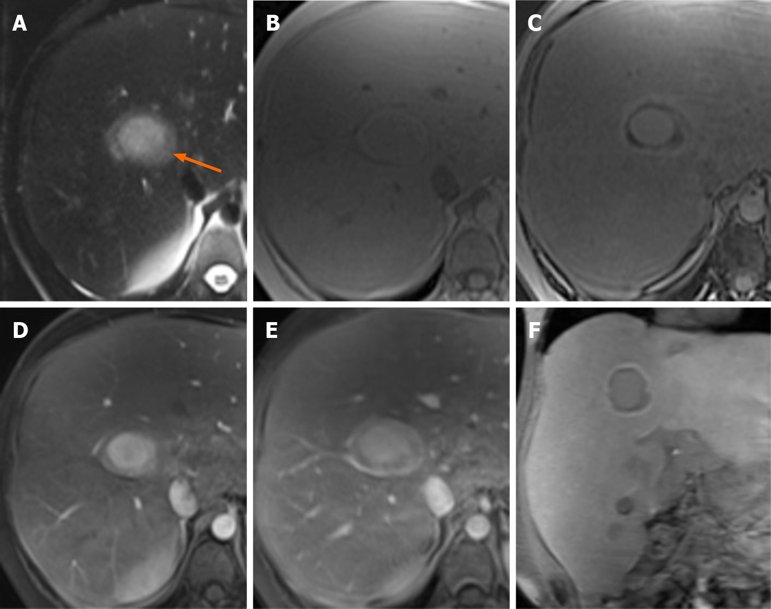
Figure 1 A 26-year-old man glycogen storage disease type I undergoing gadoxetate disodium–enhanced magnetic resonance imaging exam.
A-E: On (A) T2-weighted, (B) T1-weighted in-phase gradient-echo and (C) T1-weighted opposed phase gradient-echo magnetic resonance imaging images, the largest lesion (arrow) shows a markedly hyperintense core compared to the surrounding liver without any fat content and a slightly hyperintense peripheral thick rim that has a marked, diffuse and homogeneous fat content. T1-weighted three-dimensional spoiled gradient-echo sequences demonstrate enhancement of the lesion, that is marked in the core and mild in the periphery, during the arterial phase (D) and persistence of the enhancement in the core of the lesion during the portal venous phase (E); F: On the corresponding image, during the hepatobiliary phase, the lesion shows isointensity of the central core compared with the background liver, marked hypointensity of the peripheral thick rim and a smaller rim of contrast retention at the interface of the lesion with the liver parenchyma.
- Citation: Vernuccio F, Austin S, Meyer M, Guy CD, Kishnani PS, Marin D. "Bull’s eye” appearance of hepatocellular adenomas in patients with glycogen storage disease type I — atypical magnetic resonance imaging findings: Two case reports. World J Clin Cases 2021; 9(4): 871-877
- URL: https://www.wjgnet.com/2307-8960/full/v9/i4/871.htm
- DOI: https://dx.doi.org/10.12998/wjcc.v9.i4.871


