Copyright
©The Author(s) 2021.
World J Clin Cases. Feb 6, 2021; 9(4): 864-870
Published online Feb 6, 2021. doi: 10.12998/wjcc.v9.i4.864
Published online Feb 6, 2021. doi: 10.12998/wjcc.v9.i4.864
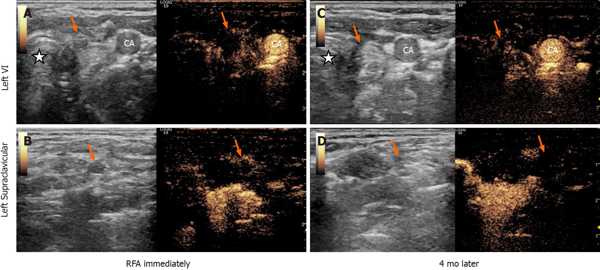
Figure 3 Images of ultrasound and contrast-enhanced ultrasound after the radiofrequency ablation.
A and B: Images captured immediately after radiofrequency ablation (RFA); C and D: Images 4 mo after RFA. The arrows point to ablated lymph nodes. Pentagram (marked with star): Trachea. CA: Carotid artery.
- Citation: Tong MY, Li HS, Che Y. Recurrent medullary thyroid carcinoma treated with percutaneous ultrasound-guided radiofrequency ablation: A case report. World J Clin Cases 2021; 9(4): 864-870
- URL: https://www.wjgnet.com/2307-8960/full/v9/i4/864.htm
- DOI: https://dx.doi.org/10.12998/wjcc.v9.i4.864


