Copyright
©The Author(s) 2021.
World J Clin Cases. Feb 6, 2021; 9(4): 854-863
Published online Feb 6, 2021. doi: 10.12998/wjcc.v9.i4.854
Published online Feb 6, 2021. doi: 10.12998/wjcc.v9.i4.854
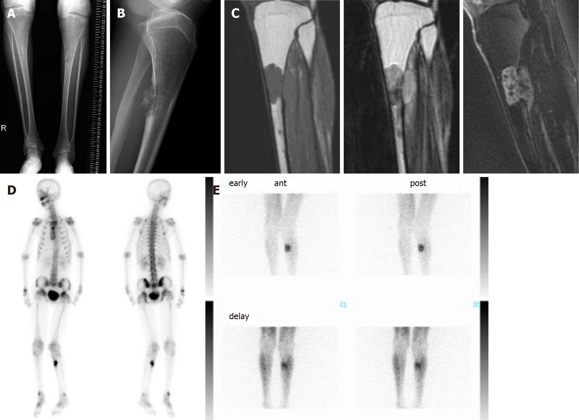
Figure 3 Image findings of bone tumor of tibia.
A and B: Bilateral tibial shafts were morphologically small in diameter and narrow in the medullary cavities. An irregular osteolytic lesion was observed in the anteroposterior view, and posterior cortical destruction and extraosseous calcified lesions were observed in the lateral view; C: Magnetic resonance imaging showed intra- and extra-osseous lesions with homogenous low intensity in T1-weighted images, low-to-high intensity in T2-weighted images, and enhanced inhomogeneity by gadolinium; D: Bone scintigraphy showed accumulation in the proximal tibia, but no other bone lesions were detected; E: Thallium scintigraphy showed high accumulation at the tumor site in the tibia.
- Citation: Hayashi K, Yamamoto N, Takeuchi A, Miwa S, Igarashi K, Araki Y, Yonezawa H, Morinaga S, Asano Y, Tsuchiya H. Long-term survival in a patient with Hutchinson-Gilford progeria syndrome and osteosarcoma: A case report. World J Clin Cases 2021; 9(4): 854-863
- URL: https://www.wjgnet.com/2307-8960/full/v9/i4/854.htm
- DOI: https://dx.doi.org/10.12998/wjcc.v9.i4.854


