Copyright
©The Author(s) 2021.
World J Clin Cases. Feb 6, 2021; 9(4): 830-837
Published online Feb 6, 2021. doi: 10.12998/wjcc.v9.i4.830
Published online Feb 6, 2021. doi: 10.12998/wjcc.v9.i4.830
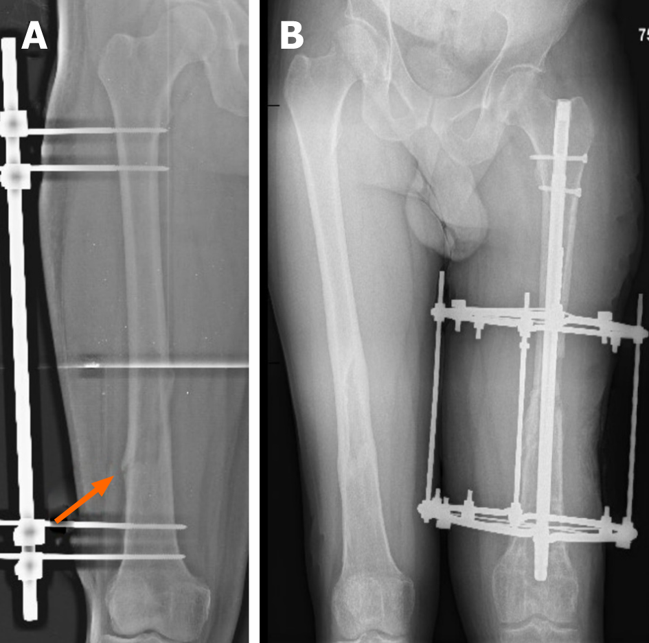
Figure 5 An X-Ray of both femurs after occurrence of pathological fractures.
A: Pathological fracture occurred in the right femur, despite previous external AO fixation. Orange arrow indicates the location of the fracture B: An X-Ray of both thighs. Right femur completely healed (after 6 mo of treatment). On the left: External fixation with Ilizarov’s apparatus, intramedullary osteosynthesis with silver-plated intramedullary nail. An X-Ray shows a significant shortening of the left limb.
- Citation: Daunaraite K, Uvarovas V, Ulevicius D, Sveikata T, Petryla G, Kurtinaitis J, Satkauskas I. Reciprocal hematogenous osteomyelitis of the femurs caused by Anaerococcus prevotii: A case report. World J Clin Cases 2021; 9(4): 830-837
- URL: https://www.wjgnet.com/2307-8960/full/v9/i4/830.htm
- DOI: https://dx.doi.org/10.12998/wjcc.v9.i4.830


