Copyright
©The Author(s) 2021.
World J Clin Cases. Feb 6, 2021; 9(4): 830-837
Published online Feb 6, 2021. doi: 10.12998/wjcc.v9.i4.830
Published online Feb 6, 2021. doi: 10.12998/wjcc.v9.i4.830
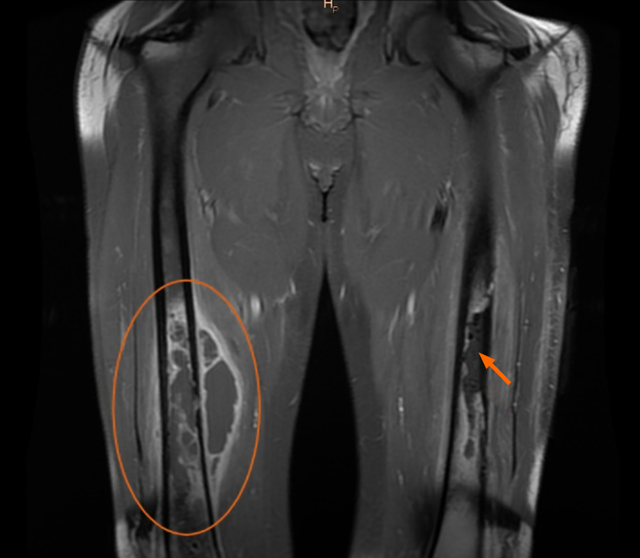
Figure 4 MRI scan of both thighs, frontal section.
The orange circle shows the abscess near the femur and in intramedullary space. The orange arrow indicates the destructive processes taking place in the left femur. Also, the photo shows the longitudinal air inserts in the interfascial space.
- Citation: Daunaraite K, Uvarovas V, Ulevicius D, Sveikata T, Petryla G, Kurtinaitis J, Satkauskas I. Reciprocal hematogenous osteomyelitis of the femurs caused by Anaerococcus prevotii: A case report. World J Clin Cases 2021; 9(4): 830-837
- URL: https://www.wjgnet.com/2307-8960/full/v9/i4/830.htm
- DOI: https://dx.doi.org/10.12998/wjcc.v9.i4.830


