Copyright
©The Author(s) 2021.
World J Clin Cases. Feb 6, 2021; 9(4): 764-773
Published online Feb 6, 2021. doi: 10.12998/wjcc.v9.i4.764
Published online Feb 6, 2021. doi: 10.12998/wjcc.v9.i4.764
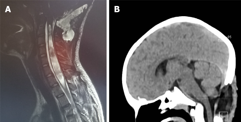
Figure 10 Two cases of craniocervical decompression.
A and B: There is a dural patch implanted in both cases. This allows an enlargement of the diameter of the dural sac at the craniocervical junction and consequently a larger space for cerebrospinal fluid circulation and rhomboencephalic structures.
- Citation: Spazzapan P, Bosnjak R, Prestor B, Velnar T. Chiari malformations in children: An overview. World J Clin Cases 2021; 9(4): 764-773
- URL: https://www.wjgnet.com/2307-8960/full/v9/i4/764.htm
- DOI: https://dx.doi.org/10.12998/wjcc.v9.i4.764


