Copyright
©The Author(s) 2021.
World J Clin Cases. Dec 26, 2021; 9(36): 11475-11481
Published online Dec 26, 2021. doi: 10.12998/wjcc.v9.i36.11475
Published online Dec 26, 2021. doi: 10.12998/wjcc.v9.i36.11475
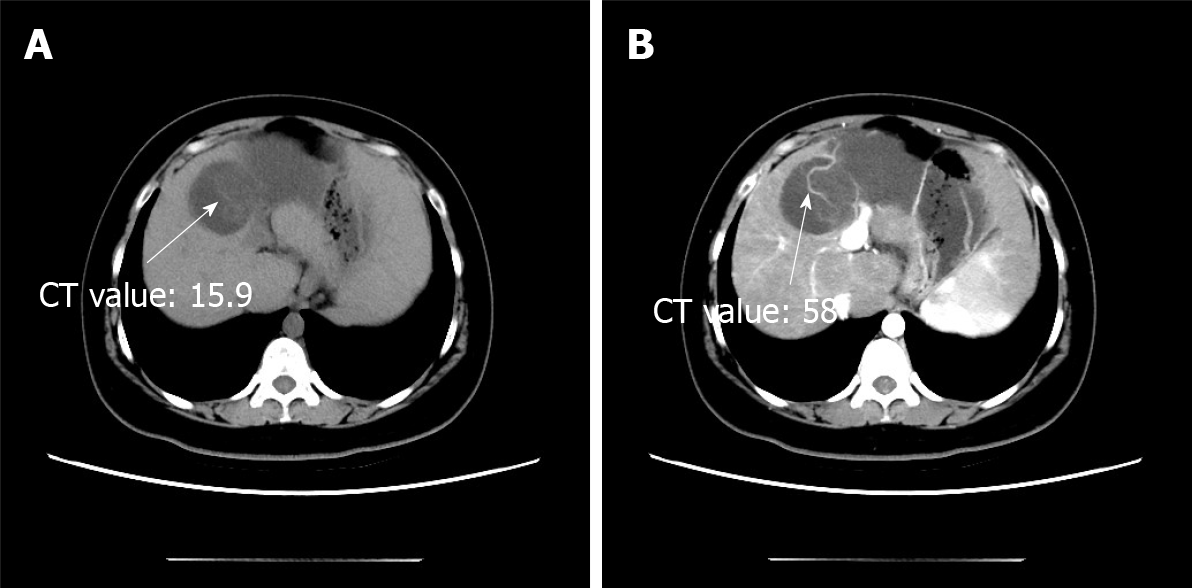
Figure 1 Computed tomography scan.
A: Non-contrast-enhanced axial computed tomography scan showing a hypodense multilocular cystic lesion; B: In the arterial phases, the cyst wall and intracystic separations are hyperdense. CT: Computed tomography.
- Citation: Yu TY, Zhang JS, Chen K, Yu AJ. Mucinous cystic neoplasm of the liver: A case report. World J Clin Cases 2021; 9(36): 11475-11481
- URL: https://www.wjgnet.com/2307-8960/full/v9/i36/11475.htm
- DOI: https://dx.doi.org/10.12998/wjcc.v9.i36.11475


