Copyright
©The Author(s) 2021.
World J Clin Cases. Dec 26, 2021; 9(36): 11467-11474
Published online Dec 26, 2021. doi: 10.12998/wjcc.v9.i36.11467
Published online Dec 26, 2021. doi: 10.12998/wjcc.v9.i36.11467
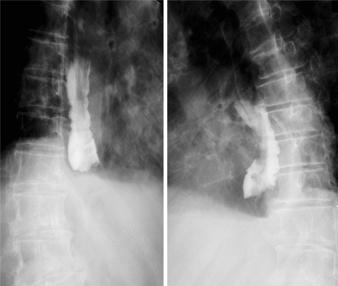
Figure 1 Esophageal X-ray barium meal angiography performed before endoscopic treatment in this case.
The lumen is obviously dilated, and an obstruction can be observed in the lower segment with only a small amount of contrast medium entering the gastric cavity, and there is no obvious extravasation.
- Citation: Hu JW, Zhao Q, Hu CY, Wu J, Lv XY, Jin XH. Rare spontaneous extensive annular intramural esophageal dissection with endoscopic treatment: A case report. World J Clin Cases 2021; 9(36): 11467-11474
- URL: https://www.wjgnet.com/2307-8960/full/v9/i36/11467.htm
- DOI: https://dx.doi.org/10.12998/wjcc.v9.i36.11467


