Copyright
©The Author(s) 2021.
World J Clin Cases. Dec 26, 2021; 9(36): 11425-11436
Published online Dec 26, 2021. doi: 10.12998/wjcc.v9.i36.11425
Published online Dec 26, 2021. doi: 10.12998/wjcc.v9.i36.11425
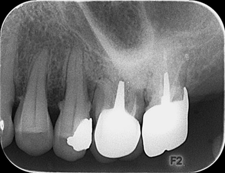
Figure 9 Radiographic image at the 2-year follow-up.
A radiograph obtained 2 years after treatment shows adequately filled canals, with no low-density area in the apical region.
- Citation: Yan YQ, Wang HL, Liu Y, Zheng TJ, Tang YP, Liu R. Three-dimensional inlay-guided endodontics applied in variant root canals: A case report and review of literature. World J Clin Cases 2021; 9(36): 11425-11436
- URL: https://www.wjgnet.com/2307-8960/full/v9/i36/11425.htm
- DOI: https://dx.doi.org/10.12998/wjcc.v9.i36.11425


