Copyright
©The Author(s) 2021.
World J Clin Cases. Dec 26, 2021; 9(36): 11425-11436
Published online Dec 26, 2021. doi: 10.12998/wjcc.v9.i36.11425
Published online Dec 26, 2021. doi: 10.12998/wjcc.v9.i36.11425
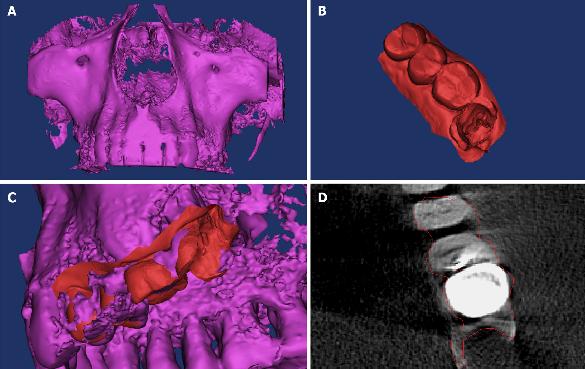
Figure 2 Establishment of a three-dimensional model of the dental hard tissue and root canal.
A: Cone-beam computed tomography (CBCT) of the patient’s maxillary teeth; B: The dentition was impressed on the working area using a three-dimensional (3D) scanner; C and D: The integrated 3D model. Red and purple parts represent the digital impression of the dentition and CBCT data, respectively.
- Citation: Yan YQ, Wang HL, Liu Y, Zheng TJ, Tang YP, Liu R. Three-dimensional inlay-guided endodontics applied in variant root canals: A case report and review of literature. World J Clin Cases 2021; 9(36): 11425-11436
- URL: https://www.wjgnet.com/2307-8960/full/v9/i36/11425.htm
- DOI: https://dx.doi.org/10.12998/wjcc.v9.i36.11425


