Copyright
©The Author(s) 2021.
World J Clin Cases. Dec 26, 2021; 9(36): 11406-11418
Published online Dec 26, 2021. doi: 10.12998/wjcc.v9.i36.11406
Published online Dec 26, 2021. doi: 10.12998/wjcc.v9.i36.11406
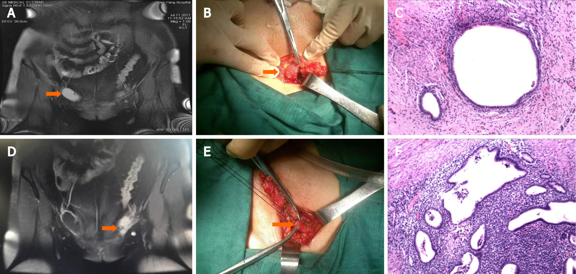
Figure 2 Magnetic resonance imaging, intraoperative findings and histological appearance of case 10.
A: The arrow points to the endometriosis lesion in the sac of the right inguinal hernia; B: The arrow points to the sac of the right inguinal hernia; C: Typical endometrial glands and stroma in the right hernia sac. Hematoxylin and eosin (H&E) staining, original magnification × 100; D and E: The arrow points to the extraperitoneal round ligament in the left inguinal region; F: Typical endometrial glands and stroma surrounded by smooth muscles. H&E, original magnification × 100.
- Citation: Li SH, Sun HZ, Li WH, Wang SZ. Inguinal endometriosis: Ten case reports and review of literature. World J Clin Cases 2021; 9(36): 11406-11418
- URL: https://www.wjgnet.com/2307-8960/full/v9/i36/11406.htm
- DOI: https://dx.doi.org/10.12998/wjcc.v9.i36.11406


