Copyright
©The Author(s) 2021.
World J Clin Cases. Dec 26, 2021; 9(36): 11237-11247
Published online Dec 26, 2021. doi: 10.12998/wjcc.v9.i36.11237
Published online Dec 26, 2021. doi: 10.12998/wjcc.v9.i36.11237
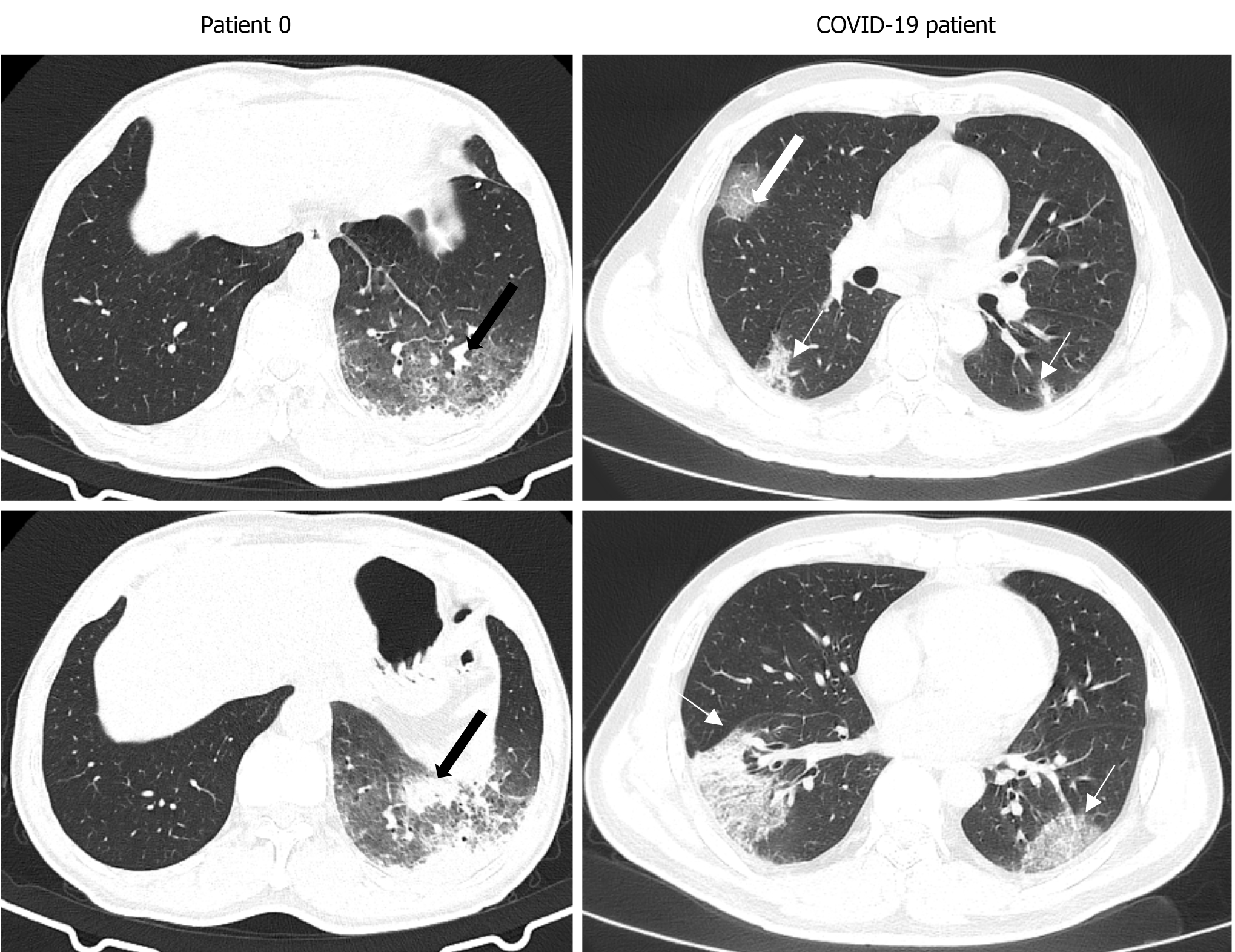
Figure 4 The computed tomography image finding of patient 0 and a case of coronavirus disease 2019.
The patient 0 have similar imaging findings (mixed ground-glass opacity and consolidation) to patients 1-4 (black thick arrow). The patients of coronavirus disease 2019 (COVID-19) presented multifocal and bilateral ground-glass opacity (white thin arrow) and vascular enlargement (white thick arrow). The margin of lesions in patients with COVID-19 is clearer. COVID-19: Coronavirus disease 2019.
- Citation: Zhao W, He L, Xie XZ, Liao X, Tong DJ, Wu SJ, Liu J. Clustering cases of Chlamydia psittaci pneumonia mimicking COVID-19 pneumonia. World J Clin Cases 2021; 9(36): 11237-11247
- URL: https://www.wjgnet.com/2307-8960/full/v9/i36/11237.htm
- DOI: https://dx.doi.org/10.12998/wjcc.v9.i36.11237


