Copyright
©The Author(s) 2021.
World J Clin Cases. Dec 26, 2021; 9(36): 11228-11236
Published online Dec 26, 2021. doi: 10.12998/wjcc.v9.i36.11228
Published online Dec 26, 2021. doi: 10.12998/wjcc.v9.i36.11228
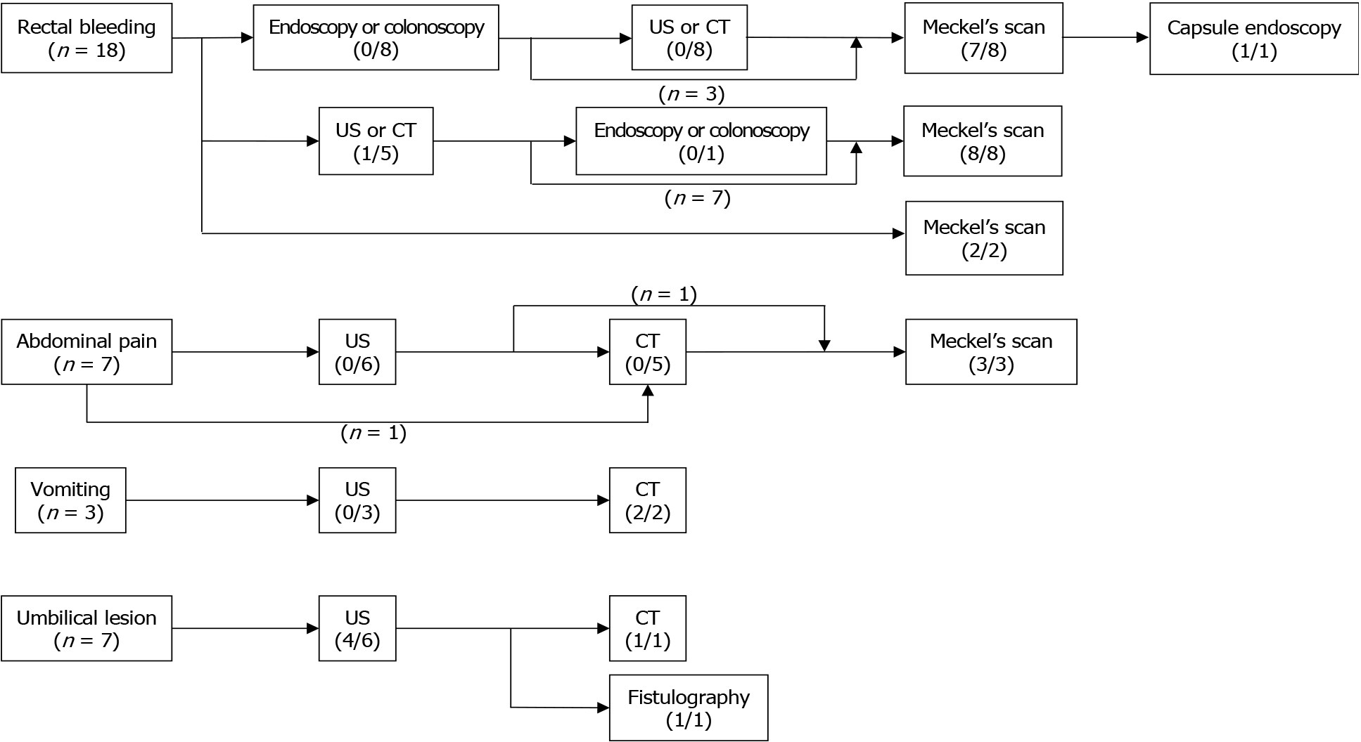
Figure 1 Flowchart of the diagnostic process for omphalomesenteric duct remnants based on chief complaints.
(1) Eighteen patients had painless rectal bleeding. Eight patients underwent endoscopy or colonoscopy initially, and all of them had negative findings; five of them underwent ultrasonography (US) or computed tomography (CT), and the remaining three patients underwent a Meckel’s scan (MS). One patient had negative findings on MS and proceeded to capsule endoscopy. Eight other patients had US or CT as the initial imaging study. Only two patients underwent a MS initially; (2) Seven patients presented with abdominal pain; of them, six patients initially underwent US and one underwent CT. All the patients had negative findings for omphalomesenteric duct remnant. Three patients underwent a MS, while four other patients underwent surgery; (3) Three patients presented with vomiting and underwent US initially, while only two patients underwent CT afterward. The patient who did not undergo CT received air reduction for intussusception followed by surgery for failed reduction; and (4) Seven patients presented with umbilical lesions. Six patients initially underwent US, while the other patient did not undergo additional imaging studies. Two patients who underwent secondary studies after US; one underwent CT and the other underwent fistulography. Figures under the modality show the number of diagnosed cases/number of performed cases. CT: Computed tomography; US: Ultrasonography.
- Citation: Kang A, Kim SH, Cho YH, Kim HY. Surgical perspectives of symptomatic omphalomesenteric duct remnants: Differences between infancy and beyond. World J Clin Cases 2021; 9(36): 11228-11236
- URL: https://www.wjgnet.com/2307-8960/full/v9/i36/11228.htm
- DOI: https://dx.doi.org/10.12998/wjcc.v9.i36.11228


