Copyright
©The Author(s) 2021.
World J Clin Cases. Dec 16, 2021; 9(35): 11115-11121
Published online Dec 16, 2021. doi: 10.12998/wjcc.v9.i35.11115
Published online Dec 16, 2021. doi: 10.12998/wjcc.v9.i35.11115
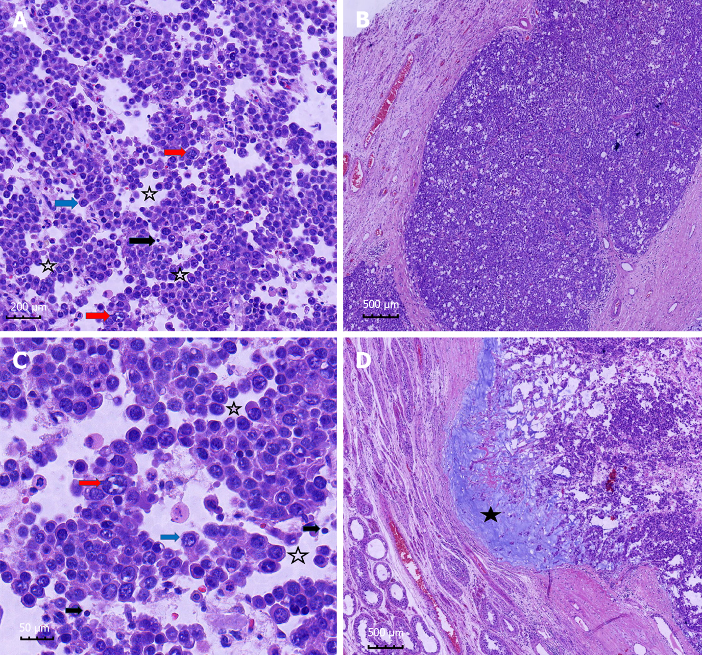
Figure 2 Histopathology findings.
A and C: Neoplastic testicular tissue with stromal edema (hollow pentagram) that produces pseudo-adenomatous change; three sizes of tumor cells were identified: Large (red arrow), small (black arrow), and medium-sized cells (blue arrow); B: Tumor cells form diffuse sheets and nests; D: Mucinous degeneration occurs in some areas (solid pentagram). Hematoxylin & eosin staining. Magnification: 100 × (A); 40 × (B and D); 400 × (C).
- Citation: Hao ML, Li CH. Spermatocytic tumor: A rare case report. World J Clin Cases 2021; 9(35): 11115-11121
- URL: https://www.wjgnet.com/2307-8960/full/v9/i35/11115.htm
- DOI: https://dx.doi.org/10.12998/wjcc.v9.i35.11115


