Copyright
©The Author(s) 2021.
World J Clin Cases. Dec 16, 2021; 9(35): 11115-11121
Published online Dec 16, 2021. doi: 10.12998/wjcc.v9.i35.11115
Published online Dec 16, 2021. doi: 10.12998/wjcc.v9.i35.11115
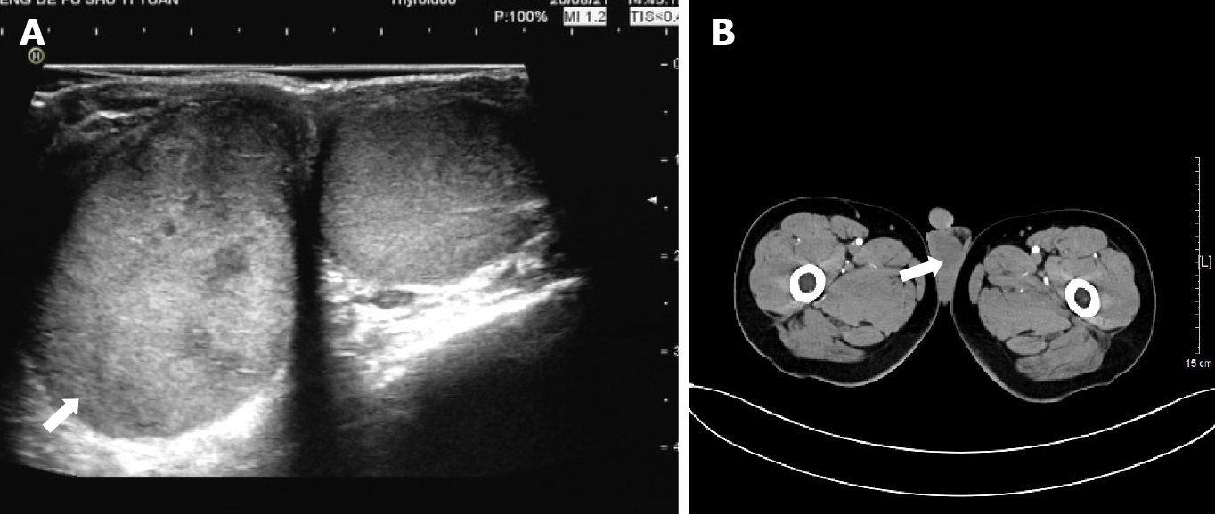
Figure 1 Ultrasound (left) and computed tomographic scans (right) of the testicles.
A: Increased volume of the right testicle, with a lesion of uneven parenchymal echo (white arrow); B: Uniform nodular change in the right testicle (white arrow).
- Citation: Hao ML, Li CH. Spermatocytic tumor: A rare case report. World J Clin Cases 2021; 9(35): 11115-11121
- URL: https://www.wjgnet.com/2307-8960/full/v9/i35/11115.htm
- DOI: https://dx.doi.org/10.12998/wjcc.v9.i35.11115


