Copyright
©The Author(s) 2021.
World J Clin Cases. Dec 16, 2021; 9(35): 11036-11042
Published online Dec 16, 2021. doi: 10.12998/wjcc.v9.i35.11036
Published online Dec 16, 2021. doi: 10.12998/wjcc.v9.i35.11036
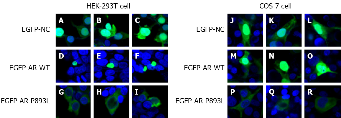
Figure 3 Subcellular localization of androgen receptor gene in human embryo kidney 293T and COS7 cells.
HEK-293T and COS7 cells were transfected with the fusion protein expression plasmid pEGFP-androgen receptor gene (AR) wild-type (WT), pEGFP-AR P893L, and the pEGFP-NC control plasmid. Twenty-four hours after transfection, cells were treated with 100 nM T. Laser confocal microscope images show that EGFP-AR WT is distributed in the nucleus (D-F, M-O), but that EGFP-AR P893L could not enter the nucleus so has a uniform distribution in the cytoplasm (G-I, P-R).
- Citation: Wang KN, Chen QQ, Zhu YL, Wang CL. Complete androgen insensitivity syndrome caused by the c.2678C>T mutation in the androgen receptor gene: A case report. World J Clin Cases 2021; 9(35): 11036-11042
- URL: https://www.wjgnet.com/2307-8960/full/v9/i35/11036.htm
- DOI: https://dx.doi.org/10.12998/wjcc.v9.i35.11036


