Copyright
©The Author(s) 2021.
World J Clin Cases. Dec 16, 2021; 9(35): 11007-11015
Published online Dec 16, 2021. doi: 10.12998/wjcc.v9.i35.11007
Published online Dec 16, 2021. doi: 10.12998/wjcc.v9.i35.11007
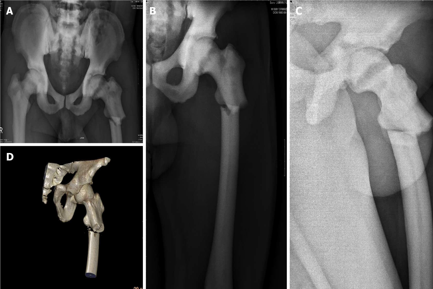
Figure 1 X-ray and three-dimensional reconstructive computed tomography of the patient after injury.
A: Antero-posterior radiograph of the pelvis showing increased bone density of pelvic bones and bilateral femurs, thickened cortical bone, and narrowed bone marrow canal; B and C: Antero-posterior and lateral preoperative radiographs of the left subtrochanteric fracture; D: 3D reconstructive computed tomography of the left subtrochanteric fracture showed discontinuous cortical bone of the left proximal femur, and separation and displacement of the bone fragments.
- Citation: Yang H, Shao GX, Du ZW, Li ZW. Treatment for subtrochanteric fracture and subsequent nonunion in an adult patient with osteopetrosis: A case report and review of the literature. World J Clin Cases 2021; 9(35): 11007-11015
- URL: https://www.wjgnet.com/2307-8960/full/v9/i35/11007.htm
- DOI: https://dx.doi.org/10.12998/wjcc.v9.i35.11007


