Copyright
©The Author(s) 2021.
World J Clin Cases. Dec 16, 2021; 9(35): 10999-11006
Published online Dec 16, 2021. doi: 10.12998/wjcc.v9.i35.10999
Published online Dec 16, 2021. doi: 10.12998/wjcc.v9.i35.10999
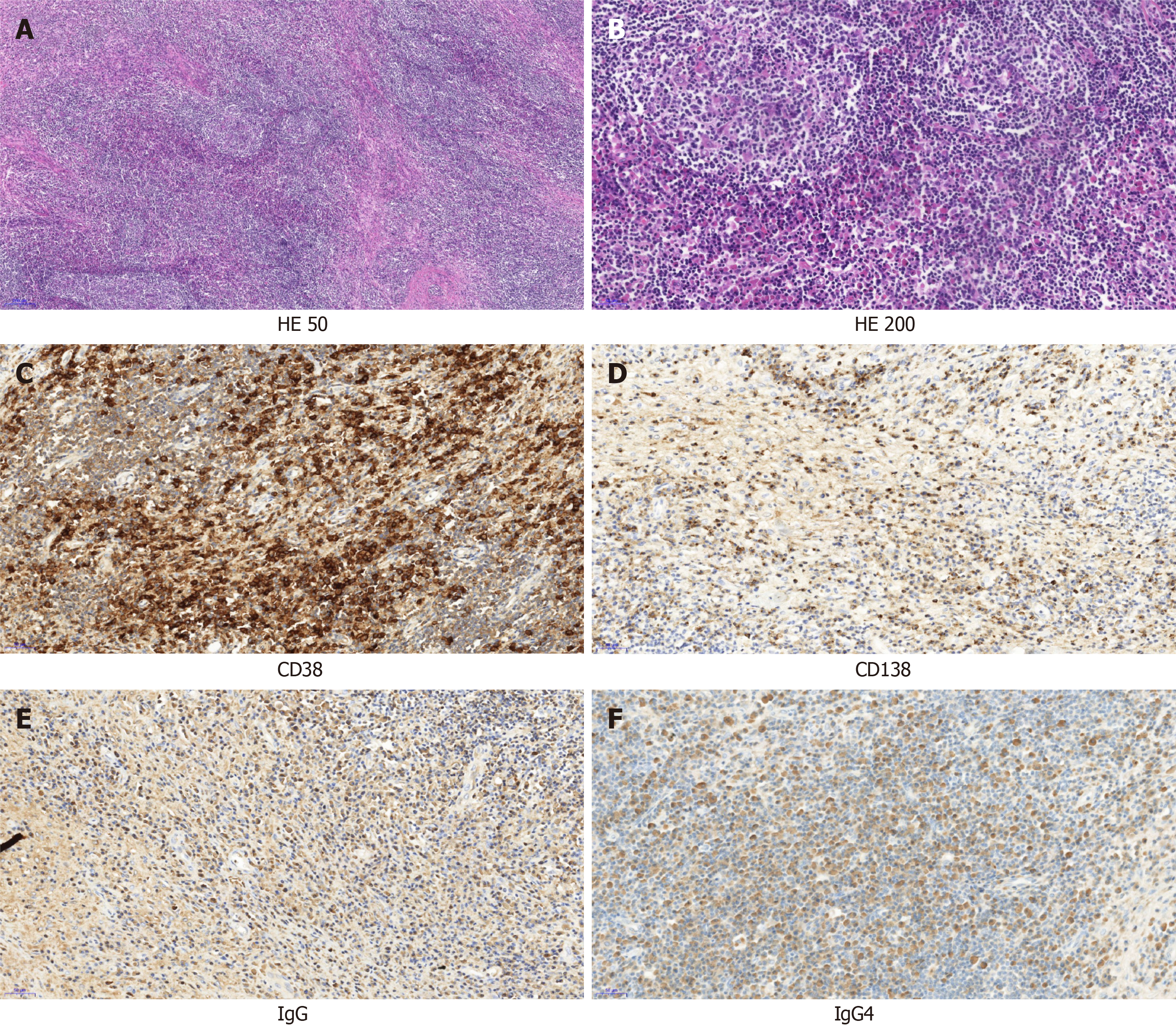
Figure 2 Pathological morphology and immunohistochemistry of orbital mass.
A: At low magnification, the histological morphology showed lymphoproliferative tissue, scattered lymphoproliferative follicles, and interstitial fibrous tissue proliferation (× 50); B: Histological morphology at high magnification showed hyperplasia of small vessels in the follicles, and a large number of plasma cells infiltrated between the follicles (× 200); C: CD38 immunohistochemical staining showed a large number of positive plasma cells in the interfollicular space; D: Immunohistochemical staining of CD138 showed a large number of positive plasma cells in the interfollicle; E: Immunoglobulin (Ig) G positive plasma cells could be seen by immunohistochemical staining; F: IgG4-positive plasma cells could be seen by immunohistochemical staining; The IgG4/IgG ratio was greater than 40%. HE: Hematoxylin and eosin stain; Ig: Immunoglobulin.
- Citation: Hao FY, Yang FX, Bian HY, Zhao X. Immunoglobulin G4-related lymph node disease with an orbital mass mimicking Castleman disease: A case report. World J Clin Cases 2021; 9(35): 10999-11006
- URL: https://www.wjgnet.com/2307-8960/full/v9/i35/10999.htm
- DOI: https://dx.doi.org/10.12998/wjcc.v9.i35.10999


