Copyright
©The Author(s) 2021.
World J Clin Cases. Dec 16, 2021; 9(35): 10828-10837
Published online Dec 16, 2021. doi: 10.12998/wjcc.v9.i35.10828
Published online Dec 16, 2021. doi: 10.12998/wjcc.v9.i35.10828
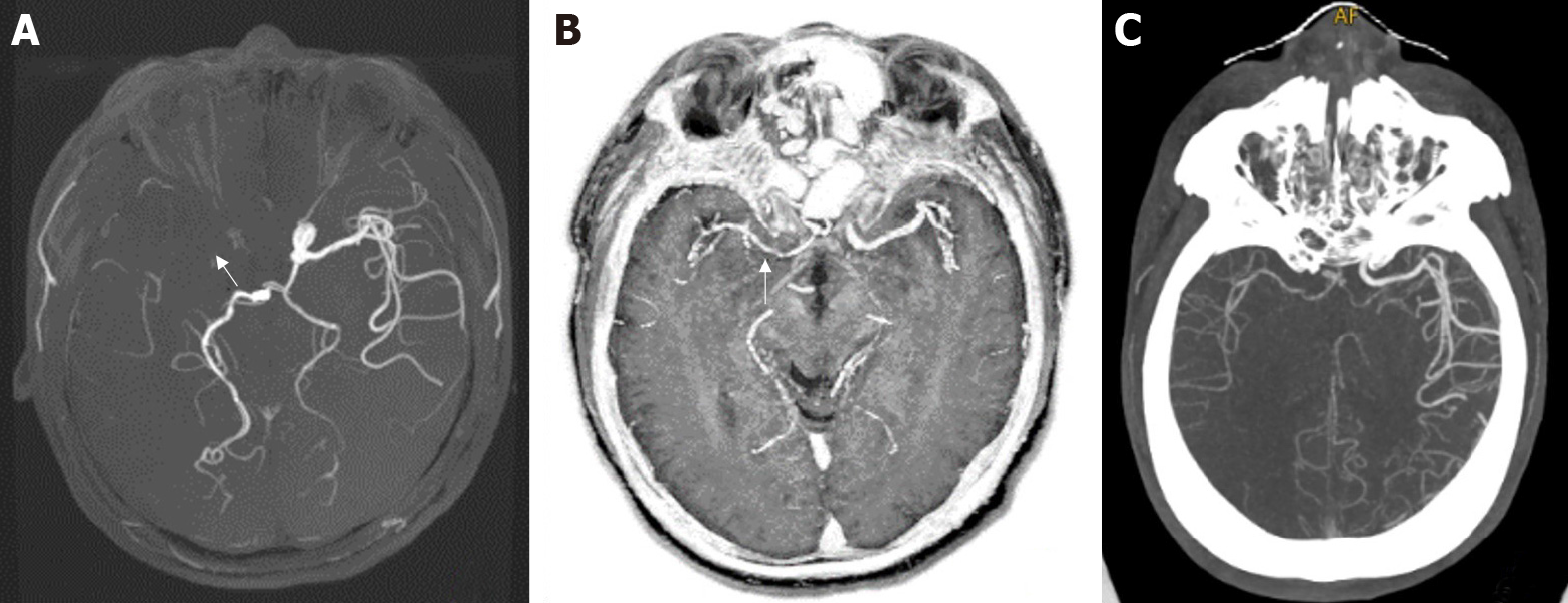
Figure 3 A 53-year-old man with right cerebral infarction.
A: The right middle cerebral artery was not shown in three-dimensional time-of-flight magnetic resonance angiography (left); B: MAGnetic resonance imaging compilation phase sensitive inversion recovery Vessel (middle); and C: Computed tomography angiography (right). Both the latter two showed mild and moderate stenosis of the local lumen of the right middle cerebral artery (arrows).
- Citation: Wang Q, Wang G, Sun Q, Sun DH. Application of MAGnetic resonance imaging compilation in acute ischemic stroke. World J Clin Cases 2021; 9(35): 10828-10837
- URL: https://www.wjgnet.com/2307-8960/full/v9/i35/10828.htm
- DOI: https://dx.doi.org/10.12998/wjcc.v9.i35.10828


