Copyright
©The Author(s) 2021.
World J Clin Cases. Dec 6, 2021; 9(34): 10733-10737
Published online Dec 6, 2021. doi: 10.12998/wjcc.v9.i34.10733
Published online Dec 6, 2021. doi: 10.12998/wjcc.v9.i34.10733
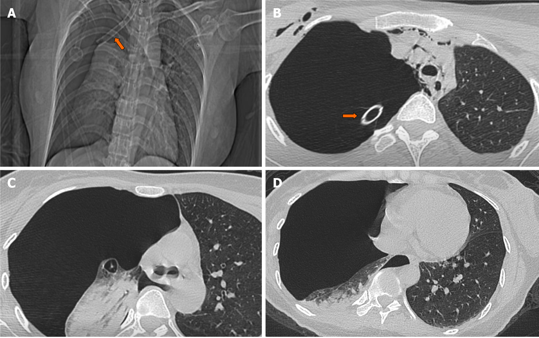
Figure 1 X-ray and computed tomography findings after insertion of endotracheal tube and right chest tube.
A and B: X ray (A) and computed tomography (CT, B) scan images revealed that there was a tracheal tube inserted into the right hemithorax (orange arrow); C and D: CT scan images showed that right pulmonary markings were absent, and right lung was condensed. The right lung was condensed by 70% and the mediastinum shifted to the right side.
- Citation: Li KX, Luo YT, Zhou L, Huang JP, Liang P. Tracheal tube misplacement in the thoracic cavity: A case report. World J Clin Cases 2021; 9(34): 10733-10737
- URL: https://www.wjgnet.com/2307-8960/full/v9/i34/10733.htm
- DOI: https://dx.doi.org/10.12998/wjcc.v9.i34.10733


