Copyright
©The Author(s) 2021.
World J Clin Cases. Dec 6, 2021; 9(34): 10681-10688
Published online Dec 6, 2021. doi: 10.12998/wjcc.v9.i34.10681
Published online Dec 6, 2021. doi: 10.12998/wjcc.v9.i34.10681
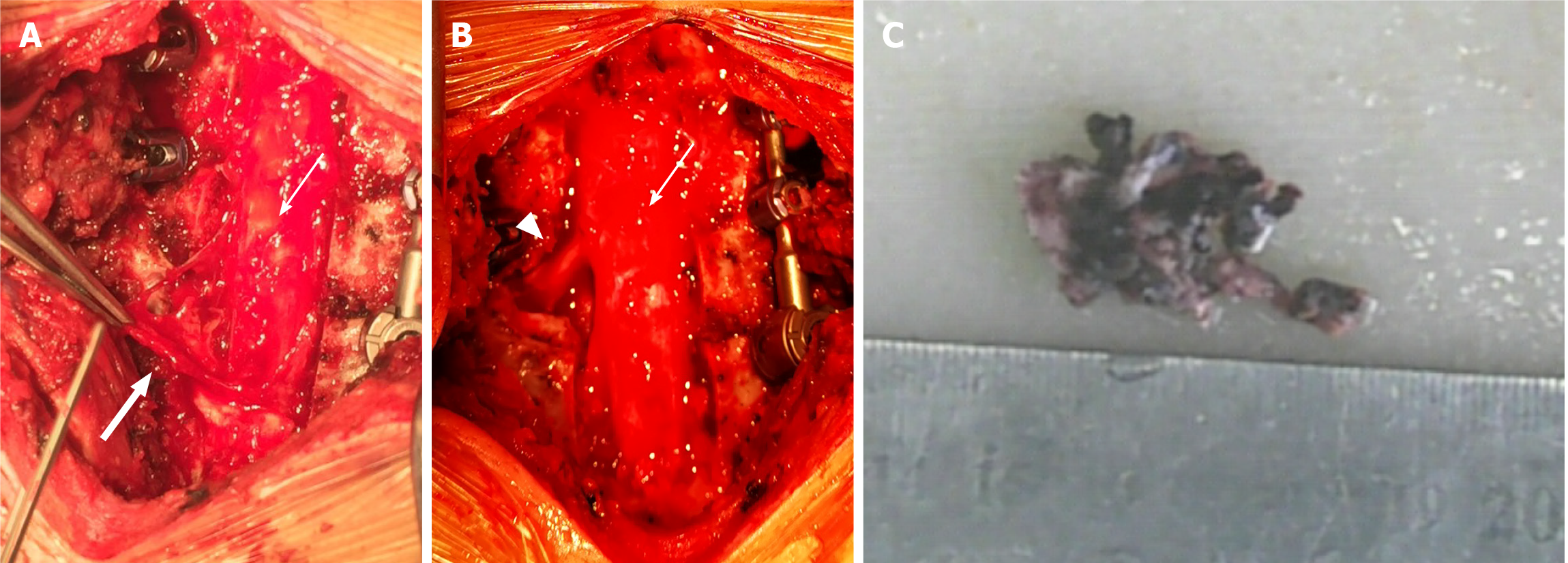
Figure 2 Intraoperative images.
A: Operative view of a dark red, nodular, highly vascularized epidural mass (thick arrow) measuring 3 cm × 1.5 cm × 1 cm compressing the left side of the spinal cord (thin arrow) after C6-T1 Laminectomy. B: View of the surgeon after complete resection of the mass and decompression of dura (thin arrow) and left C7 nerve root (triangle). C: Nodular fragment of the lesion.
- Citation: Gu HL, Zheng XQ, Zhan SQ, Chang YB. Intravascular papillary endothelial hyperplasia as a rare cause of cervicothoracic spinal cord compression: A case report. World J Clin Cases 2021; 9(34): 10681-10688
- URL: https://www.wjgnet.com/2307-8960/full/v9/i34/10681.htm
- DOI: https://dx.doi.org/10.12998/wjcc.v9.i34.10681


