Copyright
©The Author(s) 2021.
World J Clin Cases. Dec 6, 2021; 9(34): 10681-10688
Published online Dec 6, 2021. doi: 10.12998/wjcc.v9.i34.10681
Published online Dec 6, 2021. doi: 10.12998/wjcc.v9.i34.10681
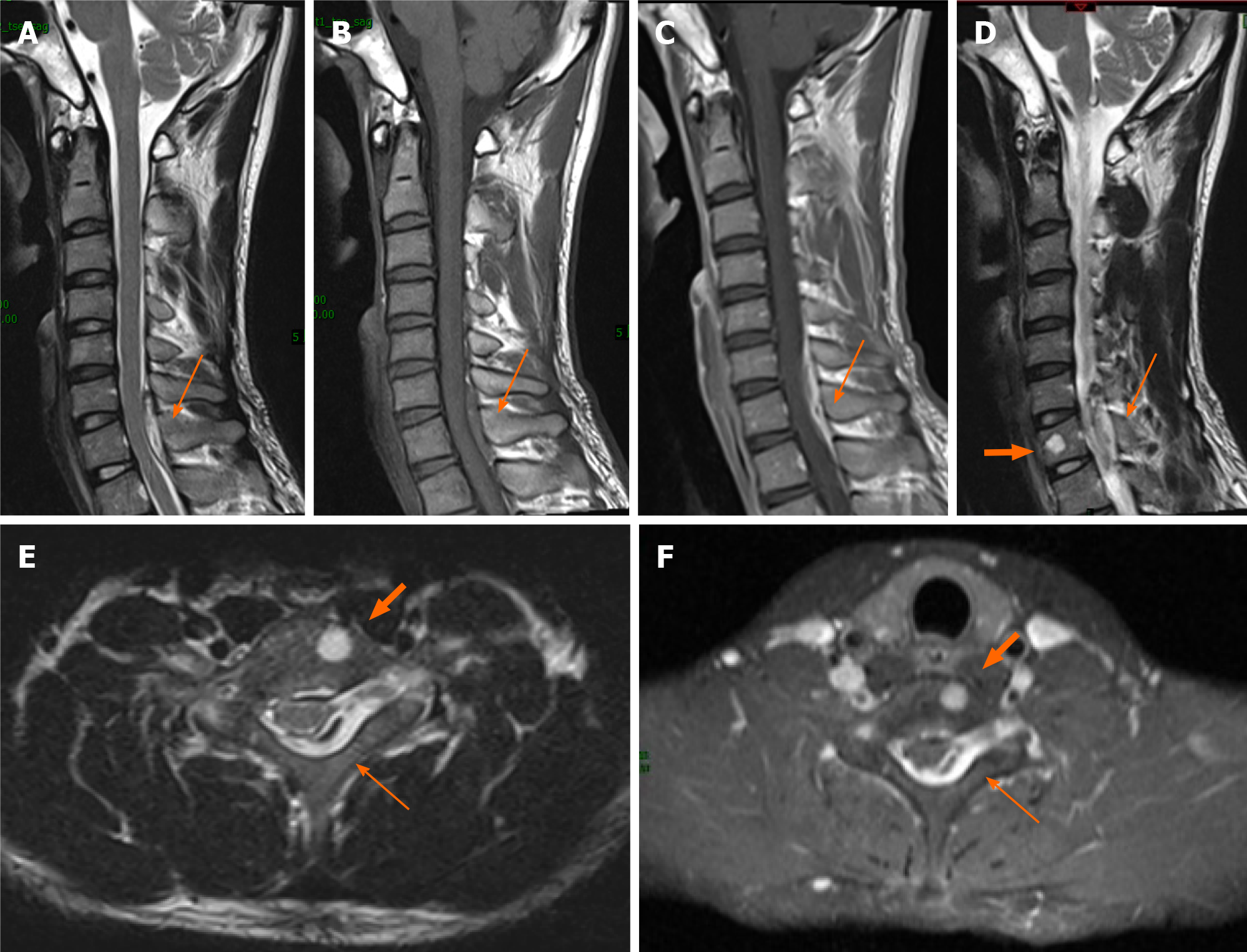
Figure 1 Preoperative magnetic resonance imaging.
A and D: Sagittal T2-weighted imaging (T2WI); B: Sagittal T1-weighted imaging (T1WI); C: Sagittal T1WI of the spine with contrast; E: Axial T2WI; F: Axial T1WI with contrast. A posterior spinal epidural mass located from C6 to T1 (thin arrow) appeared high signal intensity on T2WI sagittal and axial images, and low signal intensity on T1WI images. A gadolinium-enhanced scan reveals inhomogeneous enhancement. And a 0.5 cm × 0.5 cm × 0.6 cm-sized round tumor (thick arrow) can be seen on the left side of the C7 vertebral body; high signal intensity is observed on T2WI and homogeneous enhancement is detected on T1WI after contrast agent administration.
- Citation: Gu HL, Zheng XQ, Zhan SQ, Chang YB. Intravascular papillary endothelial hyperplasia as a rare cause of cervicothoracic spinal cord compression: A case report. World J Clin Cases 2021; 9(34): 10681-10688
- URL: https://www.wjgnet.com/2307-8960/full/v9/i34/10681.htm
- DOI: https://dx.doi.org/10.12998/wjcc.v9.i34.10681


