Copyright
©The Author(s) 2021.
World J Clin Cases. Dec 6, 2021; 9(34): 10494-10506
Published online Dec 6, 2021. doi: 10.12998/wjcc.v9.i34.10494
Published online Dec 6, 2021. doi: 10.12998/wjcc.v9.i34.10494
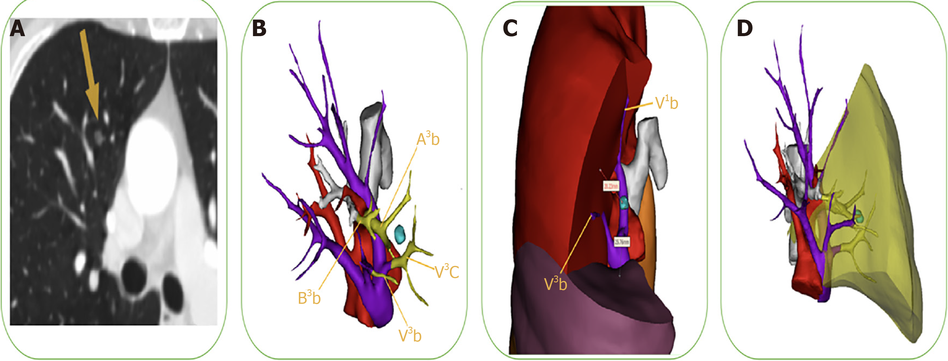
Figure 3 Identification of sufficient surgical margin and preoperative surgery simulation.
A: Chest computed tomography (CT) scan showed pure ground-glass opacity in the right upper lobe; B: We simulated surgery preoperatively, guided by a three-dimensional computed-tomography bronchography and angiography (3D-CTBA) images. The image showed that the targeted segmental structures needing to be successively resected were the V3c, A3b, B3b, and V3b; C: We precisely identified sufficient surgical margin after calculating the distance from the lesion to the predetermined cutting margin on the 3D-CTBA image. The safety margin was defined as a sphere extending at least 2 cm outside the lesion or 2 cm greater than the tumor size; D: The yellow area denotes the extent of targeted lung parenchyma RS3b. Afterward, we meticulously performed segmentectomy of the RS3b, as illustrated in Figure 4.
- Citation: Wu YJ, Shi QT, Zhang Y, Wang YL. Thoracoscopic segmentectomy and lobectomy assisted by three-dimensional computed-tomography bronchography and angiography for the treatment of primary lung cancer. World J Clin Cases 2021; 9(34): 10494-10506
- URL: https://www.wjgnet.com/2307-8960/full/v9/i34/10494.htm
- DOI: https://dx.doi.org/10.12998/wjcc.v9.i34.10494


