Copyright
©The Author(s) 2021.
World J Clin Cases. Dec 6, 2021; 9(34): 10451-10463
Published online Dec 6, 2021. doi: 10.12998/wjcc.v9.i34.10451
Published online Dec 6, 2021. doi: 10.12998/wjcc.v9.i34.10451
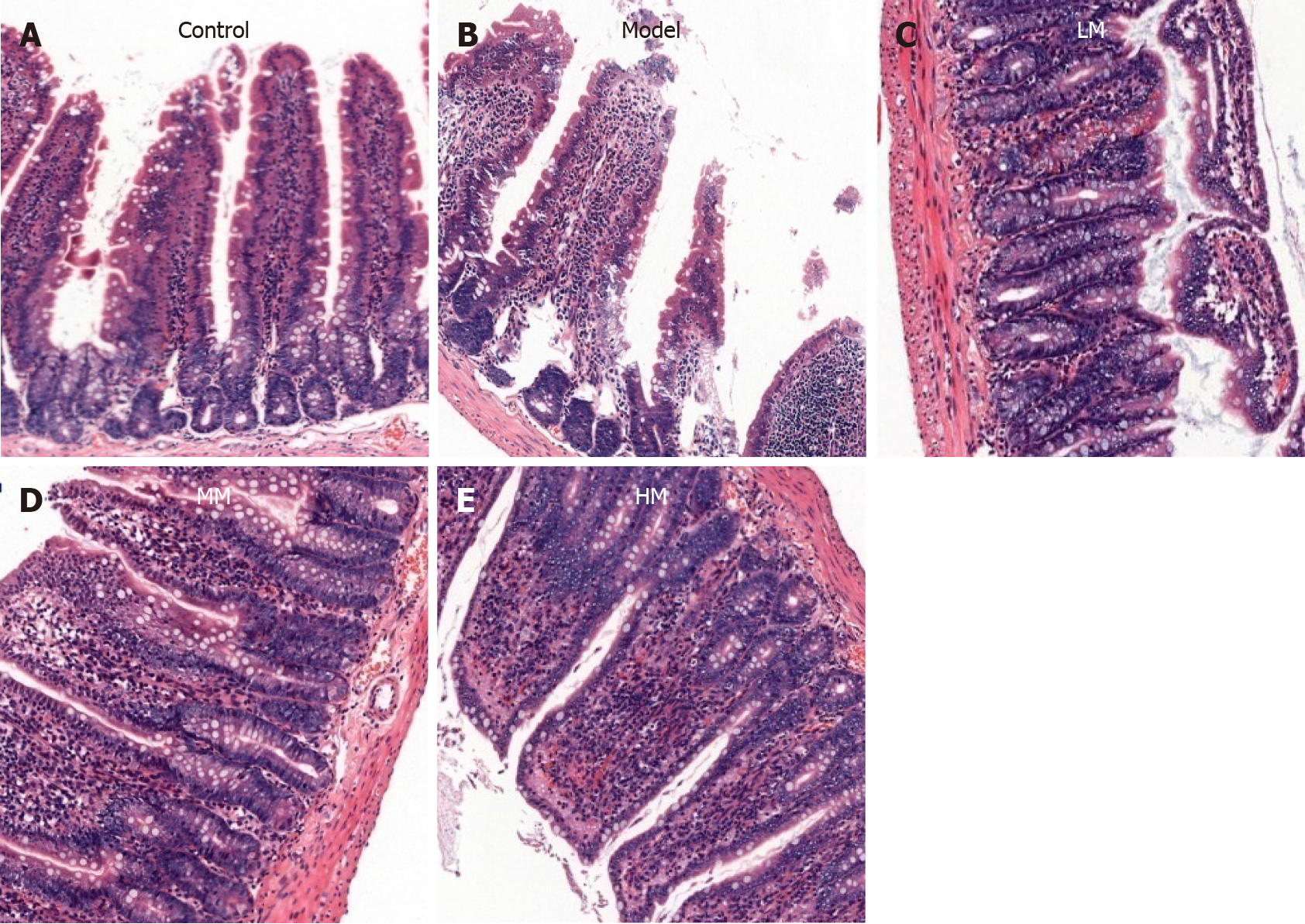
Figure 1 Hematoxylin and eosin staining of ileal tissues.
A: Control group shows orderly arranged ileal mucosal villi; B: Model group shows damaged ileal mucosal villi. The epithelial cells at the villus tips were wiped off, the subepithelial capillaries exhibited congestion, the central lacteals were expanded, the lamina propria was exposed and disintegrated, and blood capillaries were bleeding, with ulcer formation and widened intracellular tight junctions; C-E: Ileal mucosal structure in rats of the LM (C), MM (D), and HM (E) groups, respectively. The morphology of ileal mucosal villi and intestinal glands was improved to varying degrees compared to panel B. The ileal mucosal morphology was improved in panel E compared to D and in D compared to C. Magnification, × 100. Control: The control group; Model: Severe sepsis group; LM: Low-dose magnolol group; MM: Middle-dose magnolol group; HM: High-dose magnolol group.
- Citation: Mao SH, Feng DD, Wang X, Zhi YH, Lei S, Xing X, Jiang RL, Wu JN. Magnolol protects against acute gastrointestinal injury in sepsis by down-regulating regulated on activation, normal T-cell expressed and secreted. World J Clin Cases 2021; 9(34): 10451-10463
- URL: https://www.wjgnet.com/2307-8960/full/v9/i34/10451.htm
- DOI: https://dx.doi.org/10.12998/wjcc.v9.i34.10451


