Copyright
©The Author(s) 2021.
World J Clin Cases. Nov 26, 2021; 9(33): 10382-10391
Published online Nov 26, 2021. doi: 10.12998/wjcc.v9.i33.10382
Published online Nov 26, 2021. doi: 10.12998/wjcc.v9.i33.10382
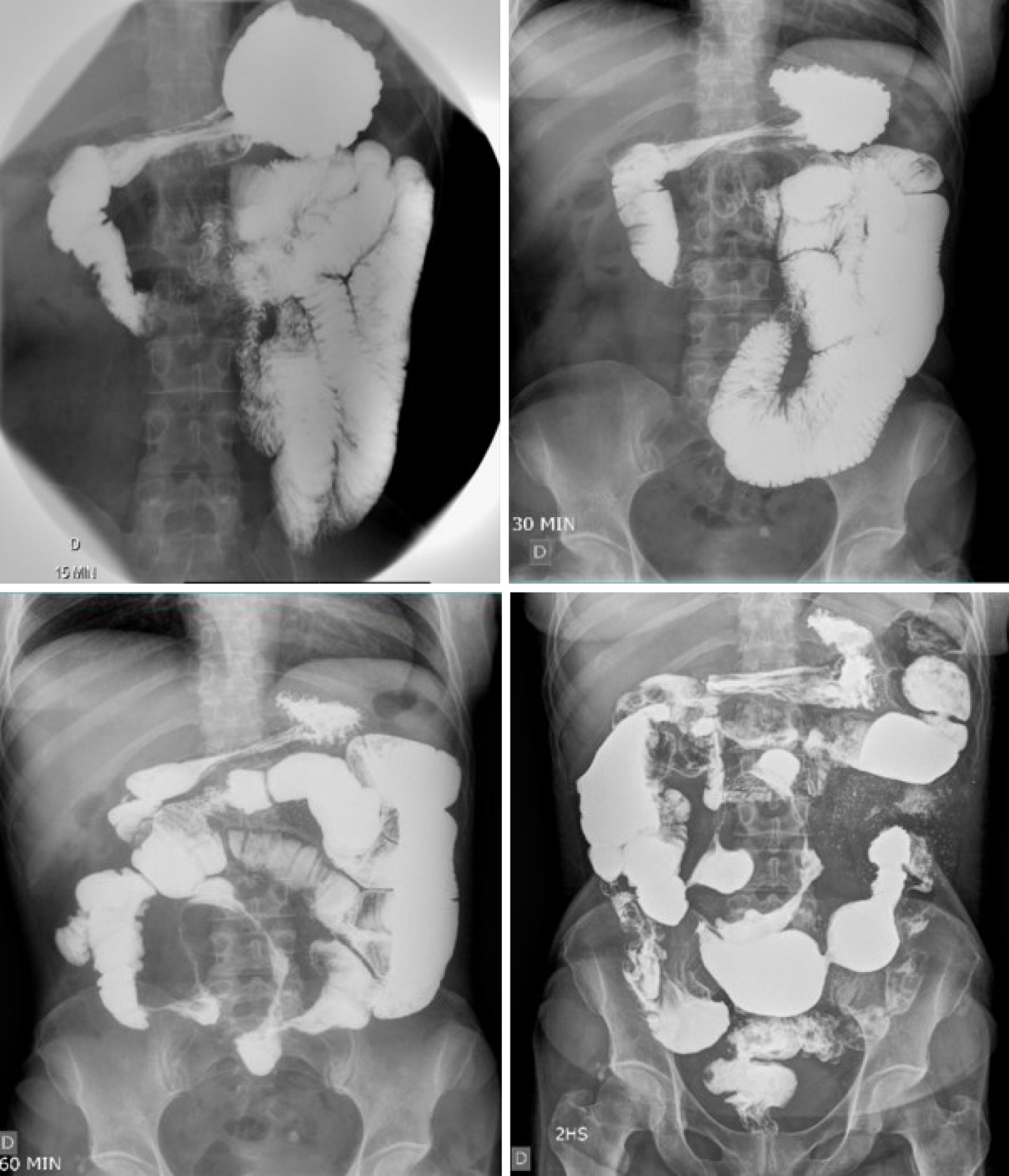
Figure 1 Small bowel follow-through showing accelerated intestinal transit.
Areas of stenosis in portions of the ileum, and the ileocecal valve with filiform aspect interspersed with areas of intestinal dilation. Presence of mucous relief irregularity with “cobblestone” images due to filiform ulcerations.
- Citation: Grillo TG, Almeida LR, Beraldo RF, Marcondes MB, Queiróz DAR, da Silva DL, Quera R, Baima JP, Saad-Hossne R, Sassaki LY. Heart failure as an adverse effect of infliximab for Crohn's disease: A case report and review of the literature. World J Clin Cases 2021; 9(33): 10382-10391
- URL: https://www.wjgnet.com/2307-8960/full/v9/i33/10382.htm
- DOI: https://dx.doi.org/10.12998/wjcc.v9.i33.10382


